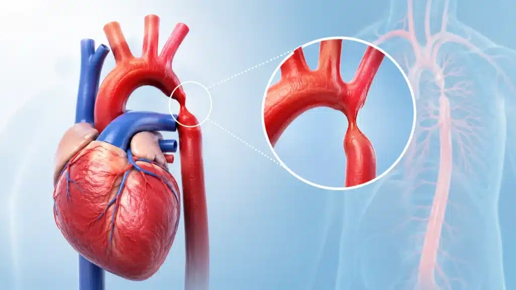
Coarctation of the aorta is a congenital heart condition in which a section of the aorta becomes narrower than normal. The aorta is the largest artery in the body. It carries oxygen-rich blood from the heart to the brain, organs, arms, legs, and the rest of the body.
When part of the aorta is narrowed, the heart must pump harder to push blood through the restricted area. This can result in high blood pressure in the arms and upper body, while blood flow to the lower body and legs may be reduced. In severe cases, particularly in newborn babies, coarctation of the aorta can cause heart failure, poor circulation, breathing difficulty, and shock soon after birth. In milder cases, the condition may not be detected until childhood, adolescence, or adulthood.
Some people with coarctation of the aorta do not have obvious symptoms for many years. They may be diagnosed after a doctor notices high blood pressure, weak pulses in the legs, a heart murmur, or a difference between blood pressure readings in the arms and legs. Because the condition affects the structure of a major blood vessel, it requires lifelong follow-up even after successful treatment.
Coarctation of the aorta, commonly called coarctation of the aorta or CoA, is a narrowing in a particular part of the aorta. The narrowing may be short and sharply defined, or it may involve a longer section of the vessel.
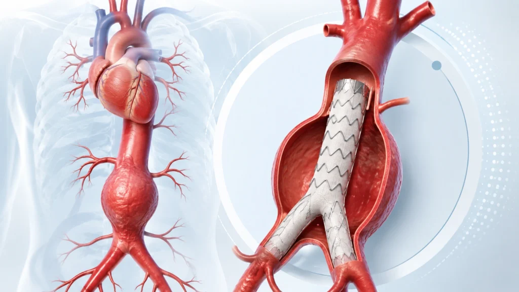
The aorta begins at the left side of the heart and travels upward before curving downward through the chest and abdomen. It gives off branches that supply blood to the brain, arms, chest, abdominal organs, and legs. In coarctation, blood flow is restricted at one point, most often in the upper part of the descending aorta.
Because the heart has to work harder to push blood through the narrowed area, the left ventricle, the main pumping chamber, may become thickened. This is called left ventricular hypertrophy. Over time, untreated coarctation can place significant strain on the heart and may lead to heart failure or persistent high blood pressure.
The condition can affect blood flow differently in each patient. Some children have severe narrowing and become critically ill when the normal fetal circulation pathway closes after birth. Others have enough blood flow through alternative vessels, called collateral vessels, and may remain stable for years.
How normal blood flow is affected:
Coarctation can be classified according to its location, appearance, timing of symptoms, and severity.
Based on location
Based on age at presentation
Based on severity
Recoarctation: Sometimes narrowing returns after previous treatment. This is called recoarctation. It may occur after surgery or catheter-based treatment and requires follow-up imaging and assessment.
Coarctation of the aorta is one of the congenital heart defects that can affect newborns and children. It may occur alone or alongside other heart abnormalities. It accounts for a significant proportion of congenital heart disease and is estimated in some medical literature at approximately three cases per 10,000 births.
The exact appearance and severity can vary widely. Some babies require urgent treatment in the first days of life, while other patients remain undiagnosed until adulthood. This variation is one reason why regular blood pressure checks and careful cardiovascular examinations are important.
The precise cause of coarctation is not always known. It develops during fetal growth and is usually considered a congenital condition. The narrowing may result from abnormal development of the aortic arch or from changes around the ductus arteriosus.
Possible causes and contributing factors
Coarctation may occur with other structural heart problems. Identifying these conditions is important because they can influence treatment and long-term follow-up.
Associated heart conditions may include:
Because coarctation is usually present from birth, the main risk factors are related to congenital development and associated genetic or cardiovascular conditions.
Severe coarctation may become apparent when the ductus arteriosus begins to close after birth. A newborn who initially appears stable may become unwell over the next few days.
Coarctation Symptoms in Infants and Children
Some children may appear healthy despite having significant narrowing. A routine examination can sometimes detect the condition through blood pressure differences or changes in pulses.
Coarctation Symptoms in Teenagers and Adults
Adults may remain undiagnosed for years, particularly when collateral blood vessels provide enough circulation to the lower body. In adulthood, high blood pressure is one of the most common presenting findings.
Coarctation can become an emergency when blood flow is severely restricted or when a serious complication develops.
Diagnosis involves a physical examination, blood pressure measurements, pulse assessment, and imaging of the heart and aorta.
Clinical examination
Echocardiography
An echocardiogram uses ultrasound to create images of the heart and major blood vessels. It can help assess:
CT angiography
CT angiography creates detailed images of the aorta and its branches. It can show:
Magnetic resonance angiography: MRI or magnetic resonance angiography can evaluate the aorta without exposing the patient to radiation. It is particularly useful for long-term surveillance in children, adolescents, and adults who need repeated imaging.
Cardiac catheterization: Cardiac catheterization involves placing a thin tube into a blood vessel and guiding it toward the heart and aorta. It can measure pressure differences and provide detailed information about the narrowed area. It may also be used as part of treatment, such as balloon angioplasty or stent placement.
Electrocardiogram: An ECG records the heart’s electrical activity. It may show signs of strain or enlargement of the left ventricle, although a normal ECG does not rule out coarctation.
Chest X-ray: A chest X-ray may show changes in the heart or aorta. In older children and adults, it may sometimes show indirect signs caused by collateral vessels, but advanced imaging is usually needed for confirmation.
Treatment depends on the patient’s age, symptoms, severity of narrowing, pressure difference, anatomy, and associated heart conditions.
The treatment goals are to:
Emergency Treatment in Newborns
A newborn with severe coarctation may require immediate medical stabilization.
Medical Management
Medicines do not permanently remove the narrowing, but they can be important before and after repair.
Surgical Treatment
Surgery may be recommended for newborns, children, or adults when the narrowing is significant or when anatomy makes catheter treatment unsuitable.
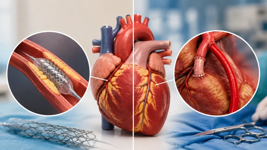
Balloon Angioplasty
Balloon angioplasty is a catheter-based treatment. A small balloon is guided to the narrowed section and inflated to widen the aorta.
Stent Placement
A stent is a tube-like mesh placed inside the narrowed part of the aorta. It supports the vessel and keeps the passage open.
Early treatment can reduce the strain caused by the narrowed aorta and protect the heart and other organs.
Benefits may include:
If coarctation remains untreated, the heart and blood vessels may be exposed to excessive pressure for years.
Recovery varies according to age, treatment type, and whether complications were present before treatment.
Recovery in children
Children may need support with:
Recovery in adults
Adults may need:
Women with repaired or unrepaired coarctation should receive specialist advice before pregnancy.
Pregnancy planning may include:
The congenital narrowing itself may not be preventable, but many long-term complications can be reduced.
Coarctation of the aorta requires more than a single procedure. Patients may need diagnosis, emergency stabilization, surgery or catheter treatment, blood pressure management, imaging, and lifelong follow-up.
It can be serious, particularly when the narrowing is severe or remains untreated. Newborns may develop heart failure or shock, while older patients may develop high blood pressure, heart strain, or aortic complications.
Some cases can be suspected during a fetal ultrasound or fetal echocardiogram, although mild narrowing may be difficult to identify before birth. A detailed assessment may be recommended when other heart abnormalities are suspected.
Medicines cannot remove the narrowed section of the aorta. They may control blood pressure, manage heart failure, or stabilize a newborn until surgery or catheter-based treatment can be performed.
Yes, recoarctation can occur after surgery or catheter treatment. Regular blood pressure checks and follow-up imaging help detect recurrence or other changes in the aorta early.
Many patients live active and fulfilling lives after successful repair. They still need lifelong cardiology follow-up because high blood pressure, aortic enlargement, or valve problems can develop later.
Many children can participate in physical activity, but the type and intensity should be guided by a cardiologist. Restrictions may be needed when blood pressure is uncontrolled or the aorta is enlarged.
Many women can have successful pregnancies after assessment and specialist monitoring. Pregnancy planning should include evaluation of the aorta, heart function, blood pressure, medicines, and associated valve disease.
Recovery varies according to the procedure and the patient’s health. Catheter treatment may allow a shorter recovery, while open surgery usually requires a longer hospital stay and gradual return to normal activity.
Coarctation of the aorta is a congenital narrowing of the body’s main artery. It can range from a mild condition discovered in adulthood to a critical emergency in a newborn. The narrowing makes the heart work harder, raises blood pressure in the upper body, and may reduce blood flow to the lower body.
Recognizing the symptoms is important. In babies, poor feeding, sweating, breathing difficulty, pale or bluish skin, extreme sleepiness, and cold limbs require urgent medical attention. In older children and adults, unexplained high blood pressure, headaches, leg cramps, cold feet, chest pain, or weak leg pulses may indicate the condition.
Treatment may involve medicines for stabilization or blood pressure control, but significant narrowing generally requires repair through surgery, balloon angioplasty, stent placement, or another specialized procedure. Early treatment can improve circulation, reduce strain on the heart, and lower the risk of serious complications.
Coarctation of the aorta (CoA) is a heart defect some babies are born with. CoA means your child’s aorta — a large artery — is pinched or narrowed in one spot.
No rows found.