
-

-

Tetralogy of Fallot

Tetralogy of Fallot

Tetralogy of Fallot

Tetralogy of Fallot: Causes, Symptoms, Diagnosis, Treatment and Long-Term Care

Tetralogy of Fallot, commonly abbreviated as TOF, is a congenital heart condition in which a baby is born with four structural abnormalities in the heart. These abnormalities change the way blood flows through the heart and lungs. As a result, the body may receive less oxygen-rich blood than it needs.

Some babies with Tetralogy of Fallot may appear slightly blue, particularly while crying, feeding, or becoming distressed. Others may have more severe symptoms immediately after birth. In some children, the condition may be detected during a pregnancy ultrasound or shortly after delivery. In other cases, the diagnosis may occur later when a child develops breathlessness, poor weight gain, tiredness, or episodes of bluish skin.

The name “tetralogy” means a group of four. In Tetralogy of Fallot, these four abnormalities usually occur together:

  • A ventricular septal defect, or VSD, which is a hole between the two lower chambers of the heart.
  • Narrowing of the pathway that carries blood from the right ventricle to the lungs.
  • An overriding aorta, in which the main artery is positioned partly over the ventricular septal defect.
  • Thickening of the right ventricular muscle, also called right ventricular hypertrophy.

What is Tetralogy of Fallot?

Tetralogy of Fallot is a heart defect present at birth. It affects the structure of the heart and changes the normal movement of blood between the heart, lungs, and the rest of the body.

In a healthy heart:

  • Oxygen-poor blood travels from the body to the right side of the heart.
  • The right ventricle pumps this blood to the lungs.
  • The lungs add oxygen to the blood.
  • Oxygen-rich blood returns to the left side of the heart.
  • The left ventricle pumps oxygen-rich blood to the body.

In Tetralogy of Fallot, the narrowed pathway from the right ventricle to the lungs makes it difficult for blood to reach the lungs. The VSD allows blood to pass between the two lower chambers. Depending on the degree of obstruction, oxygen-poor blood may move through the VSD and enter the aorta. This blood is then pumped to the body without receiving enough oxygen.

The Four Heart Defects in Tetralogy of Fallot

Ventricular septal defect

A ventricular septal defect is an opening in the wall between the right and left ventricles. In Tetralogy of Fallot, this defect is usually large.

Because of the VSD:

  • Blood can pass between the ventricles.
  • Oxygen-poor blood may move toward the aorta.
  • Oxygen-rich and oxygen-poor blood may mix.
  • The amount of oxygen delivered to the body may decrease.

Right ventricular outflow tract obstruction

The right ventricular outflow tract is the passage that carries blood from the right ventricle toward the lungs. In TOF, this pathway may be narrowed below the pulmonary valve, at the pulmonary valve, or in the pulmonary artery branches.

The obstruction may include:

  • Narrowing of the muscular outflow tract.
  • Pulmonary valve stenosis.
  • A small pulmonary valve ring.
  • Narrowing of the pulmonary arteries.
  • A combination of these changes.

Overriding aorta

The aorta is the main artery that carries blood from the heart to the body. Normally, it arises from the left ventricle. In Tetralogy of Fallot, the aorta is positioned over the ventricular septal defect and may receive blood from both ventricles.

This position makes it easier for oxygen-poor blood from the right ventricle to enter the body’s circulation. The degree of aortic override can vary from one child to another.

Right ventricular hypertrophy

The right ventricle must pump against the resistance caused by the narrowed outflow tract. Over time, the muscular wall of the right ventricle becomes thickened. This is called right ventricular hypertrophy.

The thickening is a response to increased pressure. However, it can eventually affect how efficiently the right ventricle relaxes and pumps. It is one of the structural features associated with TOF.

How Does Tetralogy of Fallot Affect Blood Flow?

The effect of Tetralogy of Fallot on blood flow depends on the degree of obstruction to the lungs.

Mild obstruction

If the pathway to the lungs is only mildly narrowed:

  • A reasonable amount of blood may still reach the lungs.
  • The child may have little or no visible cyanosis.
  • Symptoms may be mild in the newborn period.
  • The diagnosis may be made during an echocardiogram or routine evaluation.

Moderate obstruction

With moderate narrowing:

  • The child may become blue while crying or feeding.
  • Breathing may become faster during activity.
  • Feeding may take longer.
  • Weight gain may be slower than expected.
  • A heart murmur may be detected.

Severe obstruction

With severe narrowing:

  • Cyanosis may be present soon after birth.
  • The baby may have difficulty feeding.
  • Breathing may be rapid or labored.
  • The baby may become very irritable or unusually sleepy.
  • A life-threatening “Tet spell” may occur.
  • Emergency treatment may be required before surgery.

Types and Variations of Tetralogy of Fallot

Although classic TOF has four characteristic features, the anatomy can vary.

Classic Tetralogy of Fallot

This includes:

  • A large VSD.
  • Right ventricular outflow tract obstruction.
  • An overriding aorta.
  • Right ventricular hypertrophy.

TOF with pulmonary atresia

In this situation:

  • Blood cannot travel normally from the right ventricle to the lungs.
  • Lung blood flow may depend on the ductus arteriosus or other collateral vessels.
  • The baby may become severely cyanotic soon after birth.
  • Urgent specialist care is required.

TOF with absent pulmonary valve

Possible symptoms include:

  • Breathing difficulty.
  • Noisy breathing.
  • Recurrent chest infections.
  • Feeding difficulty.
  • Cyanosis.

TOF with an atrial septal defect: Some patients with TOF also have an opening between the upper chambers of the heart. This combination is sometimes described as a broader form of Fallot-related congenital heart disease.

TOF with a right aortic arch: A right-sided aortic arch may be present in some patients. This is an anatomical variation that can be identified on imaging and may influence surgical planning.

Causes of Tetralogy of Fallot

In most cases, the exact cause of Tetralogy of Fallot is not known. It develops during fetal heart formation, usually in the early weeks of pregnancy.

TOF is generally not caused by anything the parents did or did not do. Many families feel guilt after a diagnosis, but in most cases there is no single identifiable action, food, activity, or event that caused the condition.

  • Genetic changes affecting heart development.
  • Chromosomal conditions.
  • Family history of congenital heart disease.
  • Maternal diabetes that is not well controlled.
  • Certain infections during pregnancy.
  • Exposure to some medicines or harmful substances.
  • Maternal nutritional problems.
  • Alcohol or tobacco exposure during pregnancy.
  • Rare metabolic conditions.

Genetic Conditions Associated with Tetralogy of Fallot

TOF may occur alone or as part of a genetic syndrome. Possible associations include:

  • 22q11.2 deletion syndrome.
  • Down syndrome.
  • Alagille syndrome.
  • CHARGE syndrome.
  • Other chromosomal or genetic conditions.

A genetics consultation may be recommended when:

  • The child has other physical or developmental differences.
  • There is a family history of congenital heart disease.
  • Multiple birth defects are present.
  • The echocardiogram suggests an unusual heart anatomy.
  • Parents are planning another pregnancy.

Risk Factors for Tetralogy of Fallot

Most parents cannot control whether a baby develops TOF. However, some factors are associated with a higher chance of congenital heart defects.

  • Family history of congenital heart disease.
  • Maternal diabetes.
  • Certain infections during pregnancy.
  • Maternal alcohol use.
  • Smoking or exposure to tobacco.
  • Certain medicines taken during pregnancy.
  • Poorly controlled maternal metabolic conditions.
  • Older maternal age in some cases.
  • Genetic or chromosomal conditions.

Symptoms of Tetralogy of Fallot

Symptoms can appear at birth, during infancy, or later in childhood. The pattern depends on the degree of obstruction and how much oxygen reaches the body.

Common symptoms in babies

  • Bluish or grayish skin, lips, or nail beds.
  • Fast breathing.
  • Difficulty breathing during feeding.
  • Tiring quickly while feeding.
  • Sweating during feeding.
  • Poor weight gain.
  • Excessive irritability.
  • Unusual sleepiness.
  • Poor appetite.
  • Heart murmur.
  • Episodes of sudden worsening cyanosis.

Symptoms in older children

  • Breathlessness during physical activity.
  • Tiredness while playing.
  • Fainting or near-fainting.
  • Bluish lips or fingers during exertion.
  • Squatting after activity.
  • Poor exercise tolerance.
  • Delayed physical growth.
  • Clubbing of fingers in long-standing, poorly oxygenated cases.
  • Palpitations in some patients.

How is Tetralogy of Fallot Diagnosed?

Diagnosis may occur during pregnancy, soon after birth, or later in childhood.

A fetal echocardiogram may identify TOF during pregnancy. It provides a detailed view of the baby’s heart and may show:

  • A ventricular septal defect.
  • A narrowed pulmonary outflow tract.
  • An overriding aorta.
  • Abnormal pulmonary artery development.
  • Other associated heart defects.

A prenatal diagnosis allows the family and medical team to plan delivery, newborn evaluation, and treatment.

Diagnosis after birth

A newborn may be evaluated because of:

  • Low oxygen saturation.
  • Bluish color.
  • Rapid breathing.
  • Poor feeding.
  • A heart murmur.
  • Abnormal findings on routine examination.

Diagnostic tests

Pulse oximetry: A small sensor placed on the baby’s finger or foot measures oxygen saturation. Low oxygen levels may suggest a congenital heart problem, although pulse oximetry alone cannot diagnose TOF.

Echocardiogram: An echocardiogram is the main test used to diagnose Tetralogy of Fallot. It uses ultrasound waves to create images of the heart. It can show:

  • The size and location of the VSD.
  • The degree of pulmonary obstruction.
  • The position of the aorta.
  • The thickness of the right ventricle.
  • Pulmonary artery anatomy.
  • Heart function.
  • Associated defects.

Electrocardiogram: An ECG records the heart’s electrical activity. It may show signs of right ventricular strain or abnormal rhythm.

Chest X-ray: A chest X-ray may show the size and shape of the heart and the appearance of the lungs. In some cases, the heart may have a characteristic shape, although an X-ray is not sufficient for diagnosis.

Cardiac CT or MRI: Advanced imaging may be used when the anatomy is complex or when more detailed information is needed before surgery. It can help assess:

  • Pulmonary arteries.
  • Major blood vessels.
  • Surgical anatomy.
  • Collateral blood vessels.
  • Postoperative changes.

Cardiac catheterisation: Cardiac catheterisation involves passing a thin tube through a blood vessel to the heart. It may be used to measure pressures, assess blood flow, or clarify anatomy before a procedure.

Treatment Options for Tetralogy of Fallot

Treatment depends on the child’s symptoms, oxygen level, anatomy, age, and overall health. Some babies need immediate treatment, while others can be monitored for a period before surgery.

Medical stabilization

Before definitive repair, some babies may need medical support. These treatments stabilize the child but do not correct the underlying heart defects. Treatment may include:

  • Oxygen.
  • Intravenous fluids.
  • Medicines to keep the ductus arteriosus open in babies with severe pulmonary blood flow obstruction.
  • Beta-blockers to reduce the frequency or severity of Tet spells.
  • Treatment for infection or fever.
  • Support for feeding and nutrition.

Temporary or palliative procedures

Some babies are too small, medically unstable, or anatomically complex for complete repair immediately. In such cases, a temporary procedure may increase blood flow to the lungs. The goal is to improve oxygen levels and allow the baby to grow before complete repair. Possible options include:

  • A shunt procedure to connect a systemic artery to a pulmonary artery.
  • Catheter-based opening of a narrowed valve or pathway.
  • Stenting of the ductus arteriosus in selected cases.
  • Other individualized procedures based on anatomy.

Complete surgical repair

The exact operation varies from child to child. The surgeon uses echocardiography, imaging, and intraoperative findings to plan the repair. Complete repair usually involves:

  • Closing the ventricular septal defect.
  • Removing or relieving the obstruction in the right ventricular outflow tract.
  • Repairing or enlarging the pulmonary valve pathway when needed.
  • Addressing pulmonary artery abnormalities.
  • Preserving pulmonary valve function as much as possible.

When is Surgery Required?

Most children with Tetralogy of Fallot require surgical repair. The timing depends on symptoms and anatomy.

Surgery may be recommended when:

  • The baby has significant cyanosis.
  • Oxygen saturation is persistently low.
  • Tet spells occur.
  • The right ventricular outflow tract is severely obstructed.
  • Feeding and weight gain are poor.
  • The child has increasing breathlessness.
  • The anatomy is suitable for complete repair.
  • The medical team believes early repair will provide the safest outcome.

Preparing for Tetralogy of Fallot Surgery

Parents may feel anxious when surgery is recommended. The medical team explains the procedure, expected recovery, potential risks, and long-term follow-up.

Preparation may include:

  • Detailed echocardiography.
  • CT or MRI if the anatomy requires additional assessment.
  • Blood tests.
  • Evaluation of feeding and nutrition.
  • Assessment for other congenital conditions.
  • Anaesthesia consultation.
  • Discussion of blood transfusion if needed.
  • Explanation of intensive care after surgery.

Parents should inform the team about:

  • Fever or infection.
  • Feeding difficulties.
  • Previous hospital admissions.
  • Medicines being taken.
  • Allergies.
  • Family history of anaesthetic or bleeding problems.

What Happens During Surgery?

The child is placed under general anaesthesia. The surgical team uses a heart-lung bypass machine to maintain blood flow and oxygenation while the heart is repaired.

The operation may involve:

  • Opening the chest through a midline incision.
  • Closing the VSD with a patch.
  • Removing muscle that narrows the right ventricular outflow tract.
  • Repairing or enlarging the pulmonary valve and artery.
  • Reconstructing the pathway from the right ventricle to the lungs.
  • Managing associated abnormalities.

Recovery After Tetralogy of Fallot Surgery

After surgery, the child is usually cared for in a pediatric intensive care unit.

Early recovery may include:

  • Ventilator support for a period of time.
  • Monitoring of heart rate and rhythm.
  • Monitoring of oxygen levels.
  • Medicines to support heart function.
  • Pain control.
  • Fluid and electrolyte management.
  • Drainage tubes to remove fluid from the chest.
  • Gradual return to feeding.

The length of hospital stay varies. Babies with uncomplicated repair may recover faster, while children with complex anatomy may need a longer period of intensive care.

After discharge

Parents may be advised to:

  • Keep the surgical wound clean and dry.
  • Give medicines exactly as prescribed.
  • Monitor feeding, breathing, and activity.
  • Watch for fever or wound redness.
  • Attend scheduled follow-up appointments.
  • Follow instructions about bathing and physical activity.
  • Contact the hospital if the child becomes unusually tired, blue, breathless, or poorly responsive.

Possible Long-Term Problems After Surgery

Some patients remain well for many years after repair. Others may develop problems that require medicines, catheter procedures, or additional surgery.

Possible late complications

  • Pulmonary valve regurgitation.
  • Narrowing of the pulmonary valve or outflow tract.
  • Enlargement or weakness of the right ventricle.
  • Abnormal heart rhythms.
  • Reduced exercise tolerance.
  • Residual VSD.
  • Enlargement of the aorta.
  • Aortic valve leakage.
  • Heart failure in advanced cases.
  • Infective endocarditis risk in selected situations.
  • Sudden cardiac events, which are uncommon but important to monitor.

Can Children with TOF Live Normal Lives?

Many children with repaired Tetralogy of Fallot grow, learn, and participate in daily activities. Most can attend school and enjoy age-appropriate play. However, the level of physical activity should be discussed with the child’s cardiologist.

Some children may need:

  • Modified sports participation.
  • More frequent rest during physical activity.
  • School support for medical appointments.
  • Monitoring of exercise tolerance.
  • Evaluation for developmental or learning concerns.

Nutrition and Growth

Babies with TOF may have difficulty feeding because feeding requires effort and coordination. They may become tired, breathe rapidly, or sweat while feeding.

  • Taking a long time to finish a feed.
  • Frequent pauses.
  • Poor weight gain.
  • Sweating during feeding.
  • Breathing difficulty.
  • Vomiting or fatigue.
  • Reduced appetite.

Helpful nutritional support

Good nutrition is important before and after surgery because it supports healing, growth, and immune function.

  • Smaller, more frequent feeds.
  • High-calorie feeds if recommended.
  • Feeding through a tube when medically necessary.
  • Regular weight monitoring.
  • Support from a pediatric nutritionist.
  • Treatment of associated reflux or swallowing issues.

Physical Activity and Exercise

Activity recommendations vary according to the child’s repair, heart rhythm, valve function, oxygen levels, and exercise capacity.

Parents should not assume that every child with TOF needs complete physical restriction. The cardiology team can provide an individualized activity plan.

  • Light and moderate activity may be encouraged.
  • Children should be allowed to rest when tired or breathless.
  • Competitive sports may require formal assessment.
  • Sudden fainting or chest pain during exercise requires urgent evaluation.
  • Exercise testing may be used in older children and adults.

Pregnancy and Tetralogy of Fallot

Many women who had TOF repair in childhood can become pregnant, but pregnancy should be planned with specialist guidance.

Pregnancy increases the workload on the heart. Before pregnancy, a woman may need:

  • Echocardiography.
  • ECG.
  • Assessment of pulmonary valve function.
  • Evaluation of right ventricular size and function.
  • Review of medications.
  • Assessment of aortic size.
  • Consultation with an adult congenital cardiologist and obstetrician.

The risk varies considerably, so individual evaluation is essential. Pregnancy may carry greater risk when there is:

  • Severe pulmonary valve regurgitation.
  • Significant right ventricular dysfunction.
  • Severe pulmonary hypertension.
  • Serious heart rhythm problems.
  • Severe residual obstruction.
  • A markedly enlarged aorta.

When to Seek Emergency Care?

A baby with suspected TOF who becomes blue, limp, difficult to feed, or unusually sleepy needs urgent medical assessment. Parents and adults with repaired TOF should seek urgent medical help for:

  • Sudden blue or gray discoloration.
  • Severe breathing difficulty.
  • Loss of consciousness.
  • Seizure.
  • Persistent chest pain.
  • New or severe palpitations.
  • Severe weakness.
  • Repeated vomiting with cyanosis.
  • Rapid worsening of exercise tolerance.
  • High fever with unusual sleepiness.
  • Fainting during activity.

Why Choose Rama Hospital for Tetralogy of Fallot Care?

Tetralogy of Fallot requires coordinated care because it involves diagnosis, pediatric cardiology, cardiac surgery, intensive care, imaging, nutrition, rehabilitation, and long-term monitoring.

A hospital-based congenital heart programme may provide:

  • Pediatric cardiology evaluation.
  • Fetal and newborn cardiac assessment.
  • Advanced echocardiography.
  • Cardiac imaging for surgical planning.
  • Pediatric cardiac surgery.
  • Intensive care support.
  • Management of Tet spells and cyanosis.
  • Postoperative monitoring.
  • Nutrition and feeding guidance.
  • Long-term congenital heart follow-up.
  • Transition support for adolescents and adults.

Frequently Asked Questions (FAQs)

Is Tetralogy of Fallot present from birth?

Yes. Tetralogy of Fallot develops while the baby’s heart is forming during pregnancy. It may be detected before birth, shortly after delivery, or later in childhood.

Does every child with TOF look blue?

No. The degree of cyanosis depends on how severely blood flow to the lungs is obstructed. Some children have obvious bluish discoloration, while others may have mild or no visible cyanosis initially.

Can Tetralogy of Fallot be diagnosed during pregnancy?

Yes. A fetal echocardiogram may identify TOF during pregnancy. Prenatal diagnosis helps the family and medical team plan delivery, newborn stabilization, and treatment.

Does every child with TOF need surgery?

Most children require surgery, but the timing and type depend on anatomy and symptoms. Some babies need immediate repair, while others may first receive a temporary procedure or medical stabilization.

What is the difference between complete repair and a shunt?

Complete repair closes the VSD and relieves the obstruction to blood flow toward the lungs. A shunt is a temporary or palliative procedure that increases lung blood flow before complete repair.

Can a baby with TOF breastfeed?

Some babies can breastfeed, while others become tired or breathless during feeding. Smaller, frequent feeds, special feeding support, or tube feeding may be recommended based on the baby’s condition.

Is TOF surgery risky?

Heart surgery carries risks, but modern diagnosis, surgical techniques, anaesthesia, and intensive care have improved outcomes significantly. The individual risk depends on the child’s anatomy, age, health, and associated conditions.

Can Tetralogy of Fallot cause delayed growth?

Yes. Babies with significant cyanosis or feeding difficulty may gain weight slowly. Improving oxygen levels, treating the heart defect, and providing nutrition support usually help growth and development.

Conclusion

Tetralogy of Fallot is a congenital heart condition involving four related abnormalities that affect the structure and function of the heart. The condition can reduce blood flow to the lungs, lower oxygen levels, and cause cyanosis, feeding difficulty, breathlessness, poor growth, or sudden Tet spells.

Although the diagnosis can be frightening for parents, treatment has improved significantly. Medical stabilization, catheter-based procedures, temporary shunts, and complete surgical repair can help restore blood flow and improve oxygen delivery. Many children who receive appropriate treatment grow, attend school, participate in daily activities, and lead productive adult lives.
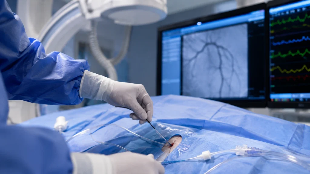
Table of Contents

Explore More

What to Know More About Tetralogy of Fallot

Find Ailments & Treatments

Search Ailments

Quickly find the information you need. Search our database to explore detailed information on various diseases and conditions, including symptoms, causes, and treatment options.

Search Treatments

Quickly find the information you need. Search our database to explore detailed information on various diseases and conditions, including symptoms, causes, and treatment options.

Recommended Reading

Video Playlist

No rows found.

Experience Healthcare the Rama Way

Emergency • OPD • Surgery • ICU Care

Need Help? Talk to our experts

Get a callback from our experts

Request a callback