Most parents never think about their newborn’s hips. The baby looks perfect. Moves normally. Cries, feeds, sleeps, all the things a baby does.
But inside, the hip joint may not have formed the way it should.
Hip dysplasia is one of those conditions that hides well. No dramatic symptoms in infancy. No obvious pain in the early years. Children grow and start walking, and the problem stays quiet, sometimes for decades, until the joint starts breaking down and the pain becomes impossible to ignore.
In India, hip dysplasia and congenital hip disorders are more common than most families realize. They affect around 1 in every 1,000 births in varying degrees of severity. They show up in teenagers with a subtle limp, in young adults with groin pain no one can explain, and in women in their 30s and 40s developing hip arthritis far too early.
The condition is treatable. Often fully correctable when caught early. But left alone, it leads to premature arthritis, chronic pain, and eventually the need for hip replacement surgery at a young age.
What is hip dysplasia?
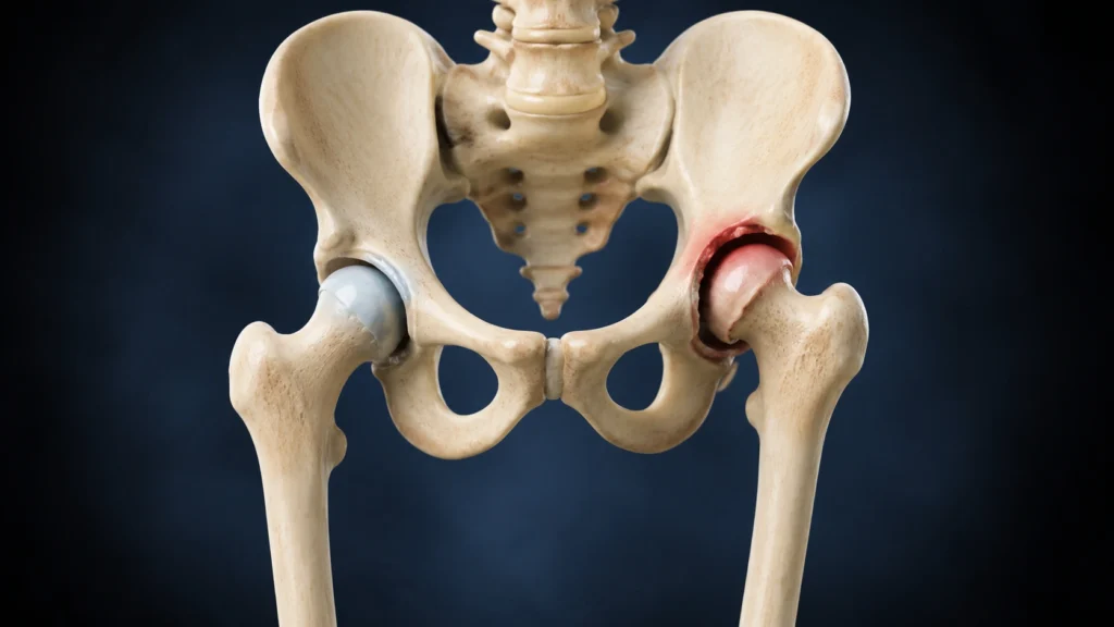
Hip dysplasia is a condition where the hip joint does not develop properly. The hip is a ball-and-socket joint; the rounded head of the femur (thigh bone) fits into a cup-shaped socket in the pelvis called the acetabulum. In a normal hip, the socket covers the ball completely, holding it securely in place.
In hip dysplasia, the socket is too shallow, too small, or incorrectly angled. The ball does not sit properly. It may be partially out of position (subluxation) or completely outside the socket (dislocation).
When this happens:
- The joint is unstable; it moves in ways it should not.
- The cartilage that lines the joint takes abnormal, concentrated stress.
- Over time, this stress destroys the cartilage.
- The result is early-onset osteoarthritis, often by the 30s or 40s.
Congenital hip disorders is the broader term covering conditions present at birth that affect hip joint development, hip dysplasia being the most common but also including complete hip dislocation, acetabular dysplasia, and coxa vara.
Types of hip dysplasia and congenital hip disorders
Not all hip dysplasia looks the same. The condition exists on a spectrum and is classified in several ways.
- Acetabular Dysplasia: The hip socket is too shallow and does not fully cover the ball of the hip joint.
- Hip Subluxation: The ball of the hip is partially out of the socket.
- Hip Dislocation (DDH): The ball of the hip is completely outside the socket from birth.
- Residual Hip Dysplasia: Hip dysplasia that was not fully corrected during childhood and continues into adulthood.
- Adult Hip Dysplasia: Hip dysplasia diagnosed later in life, often leading to early joint wear and arthritis.
- Coxa Vara: The upper thigh bone develops at an abnormal angle, affecting hip alignment.
- Femoral Anteversion: The thigh bone twists inward, causing the feet to turn inward while walking.
Developmental dysplasia of the hip: Symptoms by age group
- Newborn: Hip instability may be detected during a routine newborn examination.
- Infancy (0–6 months): Uneven skin folds or difficulty spreading the legs apart.
- Toddler / Walking Age: Limping, waddling walk, or one leg appearing shorter than the other.
- Child / Adolescent: Hip pain, reduced movement, or an unusual walking pattern.
- Adult: Groin pain, hip stiffness, or early signs of hip arthritis.
Who is most likely to have hip dysplasia?
Some children and adults are at significantly higher risk. Knowing the risk profile helps with early screening.
Newborns and Infants:
- Firstborn children: the uterus is tighter, positioning the baby in a breech more often.
- Breech presentation during delivery, the biggest single risk factor, increases risk 10 times.
- Female sex: girls are 4–5 times more commonly affected than boys.
- Family history of hip dysplasia, first-degree relatives.
- Oligohydramnios, low amniotic fluid, restricting fetal movement.
- Multiple pregnancies, twins, triplets, and restricted space in the womb.
- Large birthweight babies
- Babies swaddled with hips in an extended position (legs straight together), a practice seen in some parts of India that increases dislocation risk.
Adults:
- Young women with unexplained groin or hip pain.
- Athletes with recurrent hip injuries.
- Patients diagnosed with early hip arthritis before age 50.
- Anyone with a childhood history of treated DDH.
Causes and risk factors of hip dysplasia
- Family History: Children with a family history of hip dysplasia have a higher risk of developing the condition.
- Breech Position: Babies positioned feet-first in the womb are more likely to have abnormal hip development.
- Loose Ligaments: Hormones during pregnancy can make a baby’s hip ligaments more flexible and unstable.
- Improper Swaddling: Tight swaddling with the legs kept straight can affect healthy hip development.
- First Pregnancy: Limited space in the womb during a first pregnancy may increase the risk.
- Low Amniotic Fluid: Reduced fluid around the baby can restrict movement and affect hip formation.
- Neuromuscular Disorders: Conditions such as cerebral palsy or spina bifida can increase the risk of hip instability.
- Hip Injury: Serious trauma during childhood may affect normal hip growth and development.
Symptoms of hip dysplasia
The frustrating thing about hip dysplasia is that symptoms change completely depending on the patient’s age. What a newborn presents is nothing like what an adult experiences.
In Newborns and Infants
- Asymmetric skin folds: unequal thigh creases or buttock folds.
- Limited hip movement: one hip does not open out as far as the other.
- Clunk or click during hip examination (Ortolani/Barlow test).
- Leg length discrepancy: one leg appears shorter.
- No pain: Infants with dislocated hips are typically not in distress.
In Toddlers and Children
- Limp: the child walks with a noticeable tilt toward the affected side.
- Waddling gait: both sides affected (bilateral dysplasia).
- Toe-in or toe-out walking pattern.
- One leg appearing shorter than the other.
- Delayed walking in some children.
- Child may avoid putting weight on one leg.
In Adolescents and Young Adults
- Groin pain: deep, aching discomfort in the front of the hip.
- Pain during and after physical activity.
- Hip clicking or catching during movement.
- Stiffness after sitting for long periods.
- Difficulty with cross-legged sitting, squatting, or specific yoga positions.
- Pain that gets mistaken for a muscle strain or sports injury for years.
In Adults
- Persistent groin or hip pain: often described as deep and hard to locate.
- Pain radiating to the thigh or buttock.
- Labral tear symptoms: sharp catching pain with certain movements.
- Early hip osteoarthritis, especially in women under 50.
- Fatigue in the hip and thigh muscles.
- Limping that worsens with activity and long walks
How is hip dysplasia diagnosed?
Diagnosis depends entirely on the patient’s age. The tools and clinical tests used for a newborn are completely different from those used for an adult.
Clinical Examination in Newborns
- Ortolani Test: Checks if a dislocated hip can be gently moved back into the socket.
- Barlow Test: Checks whether a stable hip can be pushed out of the socket.
- Galeazzi Sign: Uneven knee height may indicate a hip dislocation or leg length difference.
- Skin Fold Asymmetry: Uneven thigh or buttock folds may suggest a dislocated hip.
Investigations by Age
- Ultrasound (Under 6 Months): Best test for newborns to assess hip development.
- X-ray Pelvis (After 6 Months): Shows hip bone structure and severity of dysplasia.
- MRI of the Hip: Evaluates cartilage, soft tissues, and early joint damage.
- CT Scan: Provides detailed 3D images for surgical planning.
- Arthrogram: Uses contrast dye to assess the hip joint when diagnosis is unclear.
Key Measurement Parameters
- Acetabular Index (AI): Measures how deep the hip socket is.
- Center-Edge Angle (CEA): Measures how well the socket covers the hip ball.
- Tonnis Angle: Assesses the slope of the hip socket roof.
- Lateral Centre Edge Angle: Helps diagnose hip dysplasia in older children and adults.
- Alpha Angle: Ultrasound measurement used to evaluate hip development in infants.
Treatment options for hip dysplasia
Treatment in hip dysplasia is completely age-dependent. The younger the patient, the simpler and less invasive the treatment. This is exactly why early diagnosis matters so much.
1. Pavlik Harness (0–6 Months)
The Pavlik harness is the standard first-line treatment for newborns and infants up to 6 months with DDH.
- A soft fabric harness holds the baby’s hips in a flexed, abducted position.
- This position encourages the socket to deepen naturally around the ball.
- Worn 23 hours a day initially, reduced gradually as the hip stabilises.
- Success rate of 85–95% when started before 3 months.
- Completely non-invasive, no surgery, no anesthesia.
- Duration: 6–12 weeks typically, with regular ultrasound monitoring.
2. Closed Reduction Under Anaesthesia (6–18 Months)
When the harness fails or the diagnosis is made late (6–18 months):
- The hip is gently relocated under general anesthesia.
- An arthrogram is done simultaneously to confirm correct positioning.
- A hip spica cast (plaster from waist to legs) is applied to hold the hip in position.
- Casts changed every 6–8 weeks over 3–6 months.
- Avoids open surgery in most cases when done at the right age.
3. Open Reduction Surgery
When closed reduction fails or the diagnosis is made after 18 months:
- Open surgical procedure to directly visualise and reposition the hip.
- Tight muscles and ligaments are released.
- The femoral head is placed accurately into the socket.
- A hip spica cast follows for several months.
- Often combined with osteotomy (see below) in older children.
4. Pelvic Osteotomy (Children and Adolescents)
When the socket itself is the problem, too shallow or incorrectly angled, the bone must be restructured.
- Salter Innominate Osteotomy (18 months–6 years): Repositions the hip socket to improve coverage of the femoral head.
- Triple Innominate Osteotomy (6–15 years): Allows greater correction of the hip socket in older children.
- Ganz Periacetabular Osteotomy (PAO) (Adolescents & Adults): Reshapes and repositions the hip socket while preserving the natural joint.
- Dega Osteotomy (Children with Neuromuscular Hip Dysplasia): Deepens the hip socket to provide better stability and support.
5. Periacetabular Osteotomy (PAO): The Gold Standard for Adults
The Ganz PAO is the most important surgical procedure for young adults with hip dysplasia and no severe arthritis.
- Four cuts are made around the acetabulum.
- The socket is repositioned in three dimensions.
- Fixed in corrected position with screws.
- Preserves the native hip joint completely, no replacement needed.
- Dramatically reduces joint stress and prevents or delays arthritis.
- Ideal age: 16–40 years with preserved cartilage.
- Recovery: Full weight-bearing in 6–8 weeks, full activity by 6 months.
- Long-term results: 70–80% of patients avoid hip replacement for 20+ years.
6. Femoral Osteotomy
Sometimes the problem is the femoral head angle, not just the socket.
- A cut is made in the femur to correct the angle and version of the femoral head.
- Often done alongside pelvic osteotomy for combined deformities.
- Corrects excessive anteversion (inward twisting) or varus deformity.
- Fixed with a plate and screws.
7. Hip Arthroscopy
For patients with labral tears and mild dysplasia:
- Minimally invasive keyhole surgery.
- Labral tear is repaired or reconstructed.
- Impingement is corrected.
- Best results in mild dysplasia, significant dysplasia needs osteotomy first.
- Recovery: 4–6 months for return to full activity.
8. Intra-Articular Injections
Not a cure, but helpful for managing pain while awaiting surgery or in patients not suitable for surgery:
- Corticosteroid injections reduce inflammation, provide temporary relief.
- Hyaluronic acid injections (viscosupplementation) lubricate the joint.
- PRP (Platelet-Rich Plasma) growth factors reduce inflammation and support cartilage.
- Duration of benefit: 3–6 months typically.
9. Physiotherapy and Rehabilitation
Physiotherapy plays a critical role at every stage, pre-surgery, post-surgery, and in conservative management.
Goals:
- Strengthen hip abductors, external rotators, and core muscles.
- Improve joint stability and pelvic alignment.
- Correct gait abnormalities.
- Reduce pain through targeted muscle conditioning.
Key exercises:
- Hip abductor strengthening (side-lying leg raises).
- Clamshell exercises.
- Pelvic stabilisation exercises.
- Gait retraining.
- Aquatherapy, excellent for hip dysplasia rehabilitation.
10. Bracing and Orthotic Support
- Used after closed or open reduction in infants and toddlers
- Abduction orthoses maintain corrected hip position after harness treatment
- Custom orthotics may improve gait in mild cases and reduce joint stress
11. Total Hip Replacement (THR)
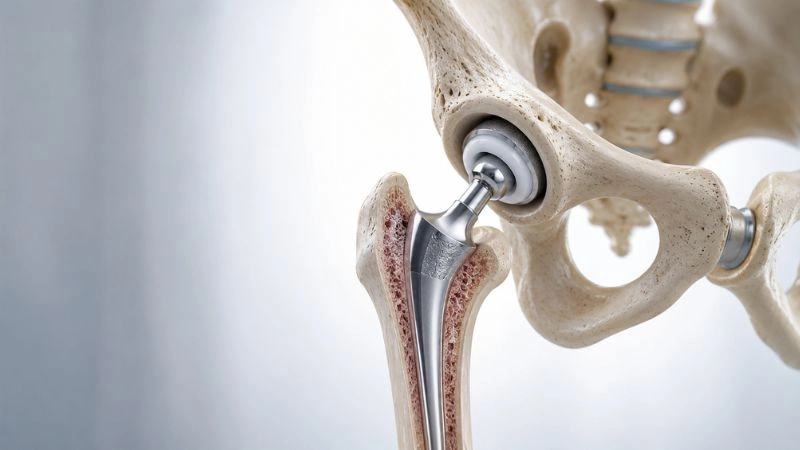
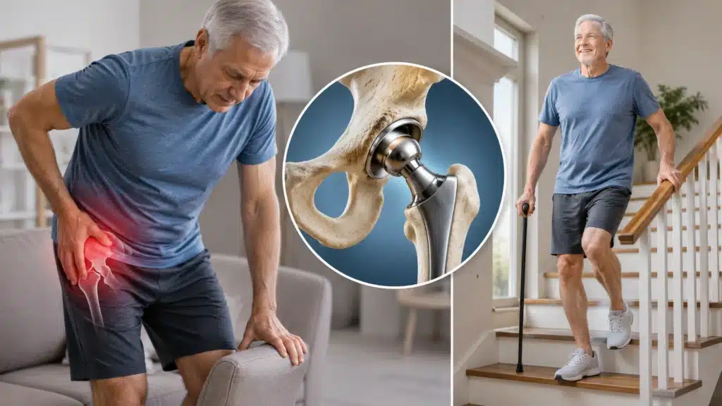
When dysplasia has progressed to end-stage arthritis, the cartilage is gone and the pain is debilitating, hip replacement is the final treatment option.
- The damaged femoral head and socket are replaced with metal, ceramic, or polyethylene implants.
- Modern THR in dysplastic hips requires specialised surgical planning.
- The socket placement is more complex due to abnormal bone anatomy.
- Dysplasia-specific implants, smaller acetabular cups, modular femoral stems, are used.
- High success rates with experienced surgeons.
- Implant survival: 15–25 years with modern implants.
- Patient returns to full independent living within 6–12 weeks.
12. Traction (Historical, Still Used in Some Cases)
- Skin or skeletal traction before surgical reduction in older children.
- Gradually stretches shortened muscles and soft tissue.
- Reduces the force needed during reduction, lowering risk of avascular necrosis.
- Used over 2–4 weeks as preparation before open reduction in select cases.
Benefits of early treatment of hip dysplasia
Starting treatment early, ideally in infancy or childhood, makes a fundamental difference to the patient’s entire life trajectory.
- Pavlik harness at birth: most infants achieve a completely normal hip with no long-term consequences.
- Avoiding hip replacement: PAO in young adults can defer or prevent replacement for 20+ years.
- Preserving cartilage: before cartilage is damaged, virtually all treatments work better.
- Normal childhood development: corrected infants walk normally, run normally, live without restrictions.
- Preventing chronic pain: adults with undiagnosed dysplasia spend years with pain, misdiagnosis, and deteriorating quality of life.
- Better surgical outcomes: operating before severe arthritis sets in dramatically improves results and recovery.
Why choose rama hospital for hip dysplasia treatment?
- Experienced specialists for newborn hip screening and DDH treatment.
- Dedicated Pavlik harness treatment with regular monitoring.
- Advanced hip-preserving surgeries for children and adults.
- Modern imaging for accurate diagnosis and surgical planning.
- Specialized hip replacement surgery for complex hip dysplasia cases.
- Comprehensive physiotherapy and rehabilitation support.
- Team-based care with orthopaedic surgeons, radiologists, and physiotherapists.
- Clear guidance on treatment plans, recovery timelines, and outcomes.
Can hip dysplasia be managed without surgery?
In some cases, yes. In others, no.
Surgery can be avoided when:
- The diagnosis is made in the newborn period and the Pavlik harness succeeds.
- The dysplasia is mild, the cartilage is healthy, and physiotherapy manages symptoms.
- The patient is elderly with limited activity demands, joint replacement may be deferred with pain management.
Surgery becomes necessary when:
- The harness fails in infancy.
- Diagnosis is made after walking age, the joint cannot self-correct at this point.
- Significant labral tear or cartilage damage has occurred.
- PAO is needed to prevent inevitable arthritis in a young adult.
- End-stage arthritis has developed.
The most important principle: do not delay assessment waiting to see if it improves on its own. In adults especially, undiagnosed dysplasia almost always progresses.
When should you to see a doctor?
For parents of newborns and infants:
- Asymmetric thigh skin folds noted at birth or during nappy changes.
- One leg appears shorter than the other.
- Hip makes a clicking sound during movement.
- Baby dislikes having one hip opened outward.
- Baby was born in breech position, arrange hip ultrasound at 4–6 weeks regardless of clinical exam.
For children and teenagers:
- Child develops a limp when beginning to walk.
- Waddling gait that persists beyond age 2.
- Child complains of hip or groin pain during activity.
- Leg length difference noticed by parent or teacher.
For adults:
- Unexplained groin pain, especially in women under 45.
- Hip pain that has been attributed to muscle strain but keeps coming back.
- Sharp catching sensation in the hip with certain movements.
- Diagnosis of early hip arthritis without obvious cause.
- History of childhood hip treatment, regular follow-up is essential.
Do not assume hip clicking in infants is normal and will resolve. Get it checked. The cost of a hip ultrasound at 6 weeks is nothing compared to the cost of surgery at 30.
What Happens If Hip Dysplasia Is Left Untreated?
Hip dysplasia does not stabilise. It progresses. The cartilage keeps wearing down until there is nothing left.
- Early Hip Arthritis – The hip joint wears out faster than normal.
- Labral Tears – The cartilage around the hip socket can get damaged.
- Bone Damage (Avascular Necrosis) – Reduced blood supply can cause part of the hip bone to die.
- Uneven Leg Length – One leg may become shorter than the other.
- Chronic Hip Pain – Ongoing joint damage can lead to persistent pain and stiffness.
- Reduced Mobility – Walking, climbing stairs, and daily activities become difficult.
- Early Hip Replacement – Severe damage may require hip replacement surgery at a younger age.
- Spine and Knee Problems – An abnormal walking pattern can put extra stress on other joints.
- Impact on Quality of Life – Long-term pain can affect work, daily routine, and emotional well-being.
Frequently Asked Questions (FAQs)
Q1. Is hip dysplasia hereditary?
Yes, genetics play a real role. If a parent or sibling has hip dysplasia, the risk in a newborn increases significantly. First-degree relatives of affected individuals should have their newborns screened with a hip ultrasound at 4–6 weeks, even if the clinical exam is normal.
Q2. My baby’s hip clicked, does that mean hip dysplasia?
Not always. Soft tissue clicking is common in newborns and often benign. However, a palpable clunk, felt during the Ortolani or Barlow test, is clinically significant and requires immediate ultrasound evaluation.
Q3. Can a child with treated DDH play sports normally?
In most cases of early-treated DDH, children grow up with completely normal hip function and participate in all sports without restriction. Children treated later or with residual dysplasia need individualised guidance, but many do play sports successfully.
Q4. I have groin pain at 32 years old. Could it be hip dysplasia?
Yes, this is actually one of the most common presentations of adult hip dysplasia. Young women with unexplained groin pain that gets worse with activity, sitting, or certain positions often have undiagnosed dysplasia. An X-ray and MRI with specific measurements can confirm it.
Q5. How long does the Pavlik harness treatment take?
Typically 6–12 weeks of full-time wear, followed by a period of part-time wear. The total duration depends on how quickly the hip stabilises on ultrasound monitoring. Most infants treated before 3 months complete treatment within 3–4 months.
Q6. Is PAO surgery very risky?
PAO is a major surgical procedure but carries acceptable risk in experienced hands. Major complications are uncommon. The main risks are blood loss, nerve injury (typically temporary), and the small risk of requiring repeat surgery.
Q7. My child was treated for DDH at birth. Does follow-up stop after treatment?
No. Children treated for DDH need orthopaedic follow-up until skeletal maturity (around age 16–18). Some residual dysplasia can develop even after successful early treatment. Regular X-rays every 1–2 years through childhood are standard practice.
Q8. Can yoga or exercises worsen hip dysplasia in adults?
Certain yoga positions, deep squats, extreme external rotation, wide-legged postures, can stress a dysplastic hip significantly. Adults with known dysplasia should work with a physiotherapist to identify which movements are safe and which should be avoided until the joint is surgically stabilised.
Conclusion
Hip dysplasia is not a rare condition. It is not a hopeless one. And it is not something families in India should be navigating alone with incomplete information.
The pattern in India is unfortunately familiar. The condition is missed at birth. The child develops a slight limp that is explained away. The teenager has hip pain after sport that is called a muscle strain. The young adult is told they have early arthritis and nothing can be done. By the time they reach a specialist, years of cartilage have been lost.
That pattern can be broken. With better awareness, routine newborn hip screening, and access to specialist orthopaedic care, most patients with hip dysplasia can live active, pain-free lives, with their own natural joint intact for as long as possible.