Skin Biopsy: Types, Procedure, Uses, Results, Risks and Recovery

A skin biopsy is a medical procedure in which a small sample of skin is removed and examined in a laboratory. It is commonly used to identify the cause of an unusual rash, persistent skin lesion, mole, lump, ulcer, infection, or suspicious growth. A skin biopsy can help determine whether a skin change is harmless, inflammatory, infectious, precancerous, or cancerous.

Many skin conditions look similar on the surface. For example, eczema, psoriasis, fungal infection, autoimmune skin disease, and some types of skin cancer may initially appear as red, scaly, itchy, or irritated patches. A clinical examination may suggest a diagnosis, but microscopic examination of skin tissue can provide more specific information.

A biopsy may also be recommended when a skin problem does not improve with routine treatment, keeps returning, changes in appearance, bleeds, develops an irregular border, or remains unexplained. In some cases, the biopsy removes the entire suspicious lesion. In others, only a small part is removed for testing.

What is a Skin Biopsy?

A skin biopsy involves removing a small amount of skin tissue for examination under a microscope. The sample is usually sent to a pathology laboratory, where a trained pathologist studies the cells and tissue structure.

The biopsy may include:

  • The outermost layer of skin, called the epidermis.
  • The deeper skin layer, called the dermis.
  • A small amount of fatty tissue below the skin, depending on the type of biopsy.
  • Part or all of a mole, lump, rash, ulcer, or other skin lesion.

What a skin biopsy can show?

A skin biopsy may help identify:

  • Skin cancer.
  • Precancerous skin changes.
  • Benign moles or growths.
  • Eczema and dermatitis.
  • Psoriasis.
  • Autoimmune blistering diseases.
  • Vasculitis.
  • Fungal or bacterial infection.
  • Certain viral infections.
  • Inflammatory skin disorders.
  • Drug-related skin reactions.
  • Unexplained ulcers or persistent rashes.
  • Disorders affecting hair follicles or sweat glands.

Why is a Skin Biopsy Done?

A dermatologist may recommend a skin biopsy when the appearance or behaviour of a skin lesion is unusual, persistent, or concerning.

Common reasons for a skin biopsy include:

  • A mole that changes in colour, size, shape, or texture.
  • A skin growth that bleeds or does not heal.
  • A lesion with an irregular border.
  • A persistent rash with no clear diagnosis.
  • A skin problem that does not respond to treatment.
  • A rapidly growing lump.
  • A painful or itchy lesion with an unusual appearance.
  • A chronic ulcer.
  • A suspected skin infection.
  • A suspected autoimmune or inflammatory disease.
  • A suspicious pigmented lesion.
  • A lesion that may be skin cancer.
  • A rash that may represent a medicine reaction.
  • A blistering disorder that requires special testing.

A biopsy may be especially useful when:

  • Several possible diagnoses appear similar.
  • The lesion is located in an unusual place.
  • The condition is recurring.
  • Treatment has failed.
  • The doctor needs to distinguish between benign and malignant growth.
  • The disease may require long-term medicines.
  • A more serious condition needs to be ruled out.

Skin Changes That May Need Evaluation

A skin biopsy may be considered for a lesion with one or more warning features.

Changes in a mole

  • Asymmetry, where one half does not look like the other.
  • Irregular or poorly defined borders.
  • Multiple colours within the same lesion.
  • Increase in size.
  • Recent change in shape or surface.
  • Itching, bleeding, or crusting.
  • A new mole appearing later in life.
  • A dark lesion that looks different from other moles.

Changes in a non-pigmented lesion

  • A sore that does not heal.
  • Repeated bleeding or crusting.
  • A shiny or pearly bump.
  • A rough, scaly patch.
  • A rapidly growing lesion.
  • A firm, persistent nodule.
  • A painful growth.
  • A lesion that changes after repeated sun exposure.

Rashes and inflammatory changes

  • A rash lasting for several weeks.
  • Severe itching with no clear cause.
  • A rash associated with fever or joint symptoms.
  • Widespread skin inflammation.
  • Recurrent blisters.
  • Thickened or scaly skin that does not improve.
  • Skin changes linked to a new medicine.
  • Unexplained purple spots or bleeding under the skin.

Different Types of Skin Biopsy

The type of biopsy depends on the depth, size, location, and suspected diagnosis of the lesion. The most common methods are shave biopsy, punch biopsy, incisional biopsy, and excisional biopsy. Other techniques, including curettage or snip biopsy, may be used in selected cases.

The choice is made to obtain enough tissue for an accurate diagnosis while limiting scarring and preserving the function and appearance of the area.ncbi.nlm.nih+1

Shave biopsy

A shave biopsy removes the upper layers of the skin using a sterile blade or similar instrument.

It may be used for:

  • Raised lesions.
  • Superficial growths.
  • Some moles.
  • Warts.
  • Certain non-melanoma skin lesions.
  • Lesions that appear limited to the upper skin layers.

Features of a shave biopsy

  • Usually performed under local anaesthesia.
  • Does not generally require stitches.
  • May leave a small shallow depression or lighter or darker mark.
  • Usually has a relatively quick healing period.
  • May not be suitable when deeper tissue is needed.

A shave biopsy may be superficial or deeper, depending on the suspected condition. The doctor chooses the depth carefully because an inadequate sample may not provide enough information.

Punch biopsy

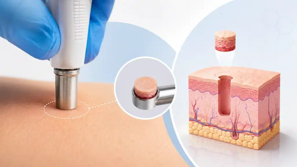
A punch biopsy uses a small circular instrument to remove a cylindrical section of skin. It usually includes the epidermis, dermis, and sometimes the upper layer of fat.

It may be used for:

  • Rashes.
  • Inflammatory skin disorders.
  • Autoimmune diseases.
  • Suspected skin cancer.
  • Pigmented lesions.
  • Chronic skin conditions.
  • Small nodules or plaques.
  • Conditions requiring full-thickness skin tissue.

Features of a punch biopsy

  • Usually performed under local anaesthesia.
  • The instrument removes a small round core of tissue.
  • One or more stitches may be required.
  • The sample is often a few millimetres wide.
  • Healing depends on the location and depth.

Punch biopsy is useful when the doctor needs to examine the full thickness of the skin rather than only its surface.

Excisional biopsy

An excisional biopsy removes the entire lesion, often along with a small margin of surrounding skin.

It may be recommended for:

  • Small suspicious moles.
  • Localised skin tumours.
  • Lesions that can be removed completely.
  • Growths where complete removal is both diagnostic and potentially therapeutic.

Features of an excisional biopsy

  • Usually performed with local anaesthesia.
  • The lesion is removed with a scalpel.
  • The wound is generally closed with stitches.
  • The removed tissue is sent for laboratory examination.
  • Further treatment may be needed if the pathology report shows that the margins are not clear.

Incisional biopsy

An incisional biopsy removes only part of a larger lesion. It is chosen when removing the entire lesion at once would be difficult, disfiguring, or medically inappropriate.

It may be used for:

  • Large lesions.
  • Extensive rashes.
  • Large tumours.
  • Lesions located near important structures.
  • Conditions requiring a representative sample before planning treatment.

Curettage biopsy

A curette is a small, spoon-shaped instrument used to scrape tissue from the surface of a lesion.

It may be considered for:

  • Certain superficial growths.
  • Some precancerous lesions.
  • Selected skin cancers.
  • Lesions where scraping is appropriate.

Curettage is not suitable for every lesion because it may not provide a complete sample or sufficient depth.

Snip biopsy

A snip biopsy uses surgical scissors to remove a small hanging skin growth, such as a skin tag.

It may be used for:

  • Skin tags.
  • Small pedunculated growths.
  • Selected superficial lesions.

The sample may be sent for testing if the appearance is unusual or the diagnosis is uncertain.

How to Prepare for a Skin Biopsy?

Most skin biopsies require little preparation, but patients should provide complete medical information before the procedure.

  • Take blood thinners.
  • Have a bleeding disorder.
  • Have diabetes.
  • Have an allergy to local anaesthetics, antiseptics, adhesives, or antibiotics.
  • Have a history of poor wound healing.
  • Have developed thick or raised scars in the past.
  • Have an artificial heart valve or other major medical condition.
  • Are pregnant or may be pregnant.
  • Have an active skin infection near the proposed biopsy site.
  • Take herbal supplements or over-the-counter medicines.

On the day of the procedure

  • Eat and drink normally unless instructed otherwise.
  • Wear comfortable clothing.
  • Avoid applying creams or makeup directly over the area.
  • Bring a list of medicines and allergies.
  • Arrange transport if the biopsy is in a difficult location or if you feel anxious.
  • Follow instructions about wound care and activity after the procedure.

What Happens During a Skin Biopsy?

A skin biopsy is usually performed in an outpatient dermatology or minor procedure setting.

Step 1: Examination and site selection

The doctor examines the lesion and identifies the most appropriate area for sampling. If the lesion is a rash, the doctor may choose an active but representative area rather than a scratched, infected, or heavily treated area.

Photographs may be taken for medical records or comparison during follow-up.

Step 2: Cleaning the skin

The skin is cleaned with an antiseptic solution. This reduces the risk of infection.

Step 3: Local anaesthesia

A small amount of local anaesthetic is injected around the biopsy site. The injection may sting or burn briefly, but the area becomes numb within a short time.

After the skin is numb, the patient should feel pressure or movement rather than sharp pain. It is important to tell the doctor if pain is felt during the procedure.

Step 4: Removing the tissue

The sample is placed in a container and sent to the pathology laboratory. The doctor uses the selected technique:

  • A blade for a shave biopsy.
  • A circular punch instrument for a punch biopsy.
  • A scalpel for an incisional or excisional biopsy.
  • A curette for a scraping biopsy.
  • Scissors for a snip biopsy.

Step 5: Closing or covering the wound

Depending on the biopsy type:

  • The area may be covered with a dressing.
  • Pressure may be applied to control bleeding.
  • A chemical or cautery method may be used for selected superficial wounds.
  • One or more stitches may be placed.
  • A larger wound may require special closure or referral for additional treatment.

Step 6: Instructions after the procedure

The procedure itself is often completed in a short period, but the exact time depends on the biopsy type and lesion location. The patient receives instructions about:

  • Keeping the dressing clean and dry.
  • Changing the dressing.
  • Applying prescribed ointment.
  • Pain relief.
  • Avoiding strenuous activity.
  • Recognising infection or abnormal bleeding.
  • Returning for stitch removal if needed.
  • Collecting or discussing the pathology report.

Is a Skin Biopsy Painful?

Most skin biopsies are performed under local anaesthesia and are generally well tolerated.

  • The anaesthetic injection may cause brief stinging.
  • The biopsy itself usually feels like pressure or pulling.
  • Mild soreness may occur after the numbness wears off.
  • Some patients experience minor bleeding or tenderness.
  • Pain can usually be managed with simple measures recommended by the doctor.

If the patient feels sharp pain during the procedure, the doctor can usually provide additional anaesthetic.

Children, highly anxious patients, and people having a biopsy in a sensitive area may need additional explanation, comfort measures, or special planning.

How Long Does a Skin Biopsy Take?

The procedure usually takes a short time, but the overall visit may be longer because of consultation, consent, preparation, wound care, and instructions.

The time depends on:

  • The size and number of lesions.
  • The type of biopsy.
  • The location of the lesion.
  • Whether stitches are needed.
  • Whether photographs or additional tests are required.
  • Whether multiple samples are taken.

What Happens to the Skin Sample?

The sample is sent to a pathology laboratory. The pathologist processes the tissue and examines it under a microscope.

  • Preserving the tissue in a special solution.
  • Embedding it in paraffin or another material.
  • Cutting very thin sections.
  • Applying stains to highlight cells and tissue structures.
  • Examining the sample under a microscope.
  • Performing special stains or immunohistochemistry when needed.
  • Sending tissue for culture if an infection is suspected.
  • Using direct immunofluorescence for selected blistering or autoimmune diseases.

The pathology report is interpreted along with the patient’s symptoms, examination findings, photographs, and medical history.

A biopsy result should not be interpreted in isolation. Sometimes the dermatologist may need to discuss the result with the pathologist or request an additional sample if the findings do not match the clinical appearance.

How Long Do Skin Biopsy Results Take?

Results may be available within several days, but the timing varies. The doctor should explain when and how the result will be communicated. Patients should not assume that a delay automatically means a serious diagnosis. Additional testing may simply be needed for accuracy. A report may take longer when:

  • Special stains are required.
  • The sample needs immunohistochemistry.
  • The tissue is sent for culture.
  • The diagnosis is complex.
  • A second pathologist’s opinion is requested.
  • Several samples are being examined.
  • The laboratory is evaluating a suspected skin cancer.

Skin Biopsy for Suspected Skin Cancer

One of the most important uses of skin biopsy is evaluating a lesion that may be skin cancer.

A biopsy can help determine:

  • Whether cancer is present.
  • The type of cancer.
  • The depth of invasion.
  • The growth pattern.
  • Whether the lesion has high-risk features.
  • Whether further surgery is needed.
  • Whether additional imaging or specialist treatment is required.

Types of skin cancer that may require biopsy

  • Basal cell carcinoma.
  • Squamous cell carcinoma.
  • Melanoma.
  • Merkel cell carcinoma.
  • Cutaneous lymphoma.
  • Rare sweat gland, hair follicle, or connective tissue tumours.

The type of biopsy is chosen carefully. For a suspicious pigmented lesion, the doctor may prefer to remove the entire lesion when practical, because the full structure can help assess melanoma.

A biopsy does not cause cancer to spread. This is a common concern, but diagnostic sampling is a standard and important part of evaluating suspicious skin lesions.

Risks and Side Effects of Skin Biopsy

Skin biopsy is generally safe, but every procedure has possible risks.

  • Mild pain or tenderness.
  • Small amount of bleeding.
  • Bruising.
  • Swelling.
  • Redness.
  • Itching as the wound heals.
  • Temporary numbness.
  • Mild scabbing.

Less common complications

  • Infection.
  • Persistent bleeding.
  • Delayed wound healing.
  • Allergy to the dressing or ointment.
  • Nerve irritation.
  • Noticeable scarring.
  • A raised scar or keloid.
  • Changes in skin colour.
  • Need for repeat biopsy.

Factors that may increase risk

  • Blood-thinning medicines.
  • Diabetes.
  • Poor circulation.
  • Smoking.
  • Weakened immunity.
  • Use of immunosuppressive medicines.
  • Previous keloid formation.
  • Infection at the biopsy site.
  • Poor nutritional status.

Recovery After Skin Biopsy

Recovery depends on the type and site of biopsy.

  • Keep the wound clean.
  • Follow instructions about keeping it dry.
  • Change the dressing as advised.
  • Apply prescribed petroleum-based ointment or medication.
  • Avoid scratching or picking the scab.
  • Protect the area from friction.
  • Avoid strenuous activity if it may reopen the wound.
  • Keep stitches dry and clean as instructed.
  • Attend the appointment for stitch removal.

Pain relief: Mild soreness is common. The doctor may recommend a suitable pain reliever. Patients should ask before taking medicines that increase bleeding, particularly if the biopsy is recent or bleeding continues.

Returning to routine: Many people return to normal daily activity the same day or the next day. Activities involving swimming, heavy sweating, contact sports, or stretching the wound may need to be delayed.

When to Contact a Doctor After a Skin Biopsy?

Most wounds heal without difficulty, but medical advice is important when healing does not appear normal.

  • Bleeding continues despite firm pressure.
  • Pain increases instead of improving.
  • The wound becomes increasingly red or warm.
  • Yellow or green discharge develops.
  • You develop fever.
  • The wound opens.
  • The dressing causes significant itching, swelling, or rash.
  • The area becomes numb or unusually sensitive.
  • Stitches break or come out early.
  • You are concerned about the appearance of the wound.

Why Choose Rama Hospital for Skin Biopsy?

A skin biopsy is a small procedure, but its accuracy depends on appropriate site selection, technique, tissue handling, and pathology interpretation. Hospital-based dermatology care can provide coordinated diagnosis and treatment when a lesion is complex or potentially serious.

  • Dermatology evaluation by experienced specialists.
  • Selection of the appropriate biopsy technique.
  • Careful assessment of suspicious moles, rashes, ulcers, and growths.
  • Local anaesthesia and outpatient procedure support.
  • Proper handling and laboratory processing of tissue samples.
  • Pathology interpretation and clinical correlation.
  • Follow-up for wound healing and biopsy results.
  • Further treatment planning for cancer, infection, or inflammatory disease.
  • Referral to oncology, surgery, rheumatology, or other specialists when required.

Frequently Asked Questions (FAQs)

What are the main types of skin biopsy?

The main types include shave, punch, incisional, and excisional biopsy. The dermatologist selects the technique based on the lesion’s size, depth, location, and suspected diagnosis.

Does a skin biopsy always mean cancer?

No. A biopsy is used to identify many conditions, including eczema, psoriasis, infections, benign moles, autoimmune diseases, and cancer. It is a diagnostic step, not a confirmation that cancer is present.

How long does a skin biopsy take?

A small biopsy is usually completed in a short outpatient visit. The total time depends on the type of biopsy, the number of lesions, the location, and whether stitches are required.

Will I need stitches after a skin biopsy?

Stitches may be needed after a punch, incisional, or excisional biopsy. Shave biopsies and some superficial procedures usually heal with a dressing and do not require stitches.

How long do biopsy results take?

Results may be available within several days, but complex cases can take longer. Special stains, immunofluorescence, cultures, or expert review may be required before the final report is issued.

Will a skin biopsy leave a scar?

A small scar or change in skin colour may remain after a biopsy. The appearance depends on the technique, location, wound size, healing process, and individual tendency to form raised scars.

Should I stop blood thinners before a biopsy?

Do not stop blood thinners without medical advice. Inform your doctor about every blood-thinning medicine so the procedure can be planned safely.

Conclusion

A skin biopsy is an important diagnostic procedure that helps doctors understand unusual or persistent skin changes. It may be recommended for a suspicious mole, changing growth, unexplained rash, chronic ulcer, infection, autoimmune condition, or possible skin cancer. The procedure is usually quick, performed under local anaesthesia, and well tolerated.

The type of biopsy depends on the lesion. A shave biopsy removes superficial tissue, a punch biopsy obtains a deeper cylindrical sample, an incisional biopsy removes part of a larger lesion, and an excisional biopsy removes the entire lesion when appropriate. The tissue is examined by a pathologist, and the result helps guide further treatment.

Most patients recover quickly, although mild soreness, bleeding, scabbing, and scarring may occur. Proper wound care and follow-up are important. Patients should contact a doctor if they develop increasing pain, redness, swelling, pus, fever, persistent bleeding, or wound separation.
Table of Contents

Explore More

What to Know More About Skin biopsy

Find Ailments & Treatments

Search Ailments

Quickly find the information you need. Search our database to explore detailed information on various diseases and conditions, including symptoms, causes, and treatment options.

Search Treatments

Quickly find the information you need. Search our database to explore detailed information on various diseases and conditions, including symptoms, causes, and treatment options.

Recommended Reading

Video Playlist

No rows found.

Experience Healthcare the Rama Way

Emergency • OPD • Surgery • ICU Care

Need Help? Talk to our experts

Get a callback from our experts

Request a callback