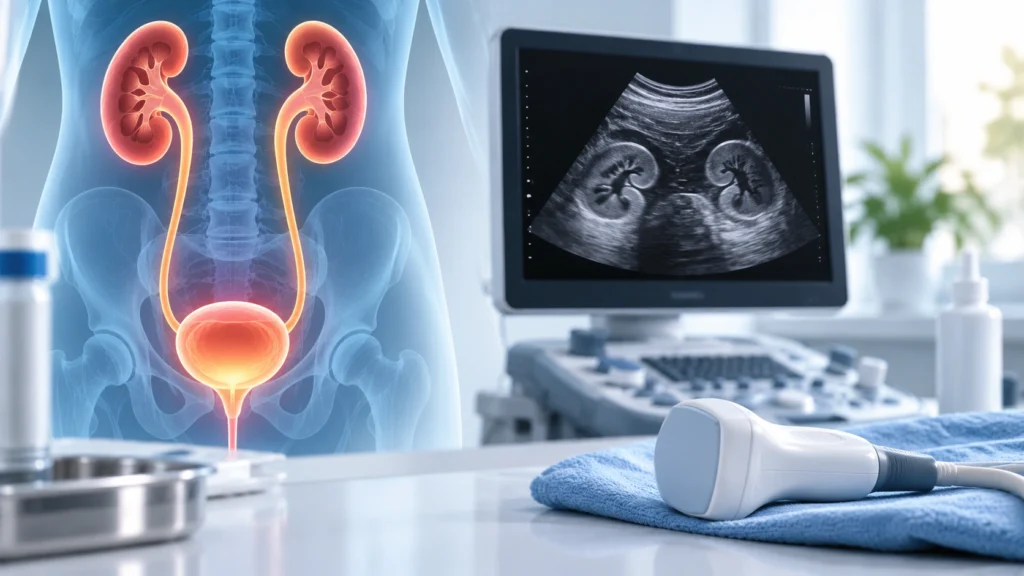
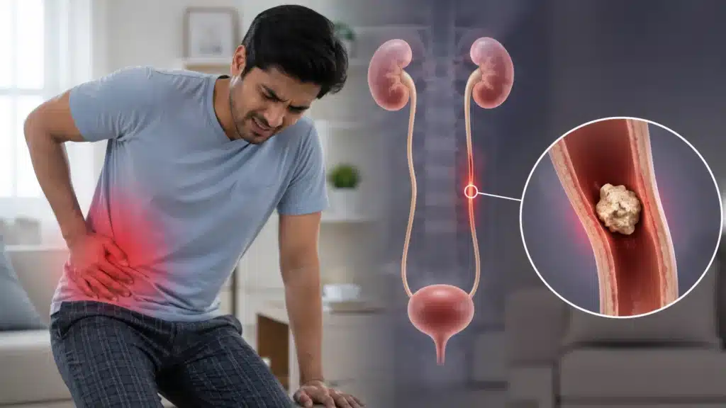
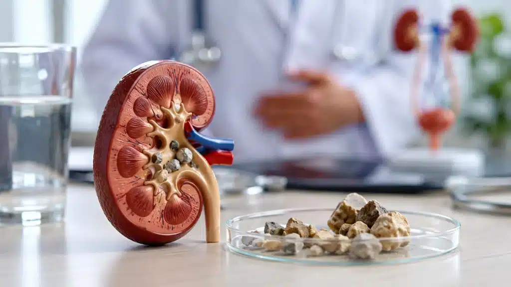
Ultrasound KUB is a simple, non-invasive imaging test that examines the kidneys, ureters, and bladder. KUB stands for Kidney, Ureter, and Bladder. This scan uses sound waves to create real-time images of the urinary system without radiation, needles, or contrast dye in most cases. It is one of the most commonly advised tests for patients with urinary symptoms, abdominal pain, suspected stones, infection, or kidney-related problems.
Many people are advised an Ultrasound KUB when they have flank pain, burning urination, blood in urine, recurrent urinary infections, swelling in the legs, or high blood pressure. The test helps doctors see the size, shape, and structure of the kidneys, check for stones or blockages, assess the bladder, and look for any abnormal growths or fluid collection. Because it is safe, painless, and widely available, it is often the first imaging test chosen for urinary complaints.
What is Ultrasound KUB?
Ultrasound KUB is an ultrasound scan focused on the urinary system. It uses a handheld device called a transducer that sends sound waves into the body. These waves bounce back from internal organs and are converted into images on a screen. The radiologist studies these images to assess the kidneys, ureters, and bladder.
The test does not use X-rays or radiation. It is safe for most people, including children and pregnant women when medically indicated. It is usually done as an outpatient procedure and takes only a short time. The patient lies on an examination table while the sonographer moves the transducer over the abdomen and lower belly to capture images from different angles.
Ultrasound KUB is different from a whole abdominal ultrasound, which also looks at the liver, gallbladder, pancreas, and spleen. KUB focuses specifically on the urinary tract, although in practice, some centers may include a quick look at related structures depending on the clinical question.
Why is Ultrasound KUB Done?
Ultrasound KUB is advised for many urinary and abdominal symptoms. It helps identify structural problems, blockages, stones, infections, and other abnormalities.
Common reasons for Ultrasound KUB
- Persistent or recurrent pain in the flank, lower abdomen, or back.
- Burning or pain during urination.
- Frequent urination or urgency.
- Blood in urine or dark-colored urine.
- Recurrent urinary tract infections.
- Swelling in legs, feet, or around the eyes.
- High blood pressure, especially if kidney cause is suspected.
- Abnormal urine or blood test related to kidney function.
- Suspected kidney stones or previous history of stones.
- Follow-up of known kidney cysts, masses, or abnormalities.
- Difficulty passing urine or incomplete emptying.
- Evaluation before and after urology procedures.
- Assessment of bladder size and wall thickness.
- Screening in patients with chronic kidney disease.
What the doctor wants to know?
- Whether the kidneys are normal in size and shape.
- Whether there are stones, cysts, or masses.
- Whether there is any blockage or swelling of the kidney.
- Whether the bladder is normal and emptying properly.
- Whether there is fluid collection or infection signs.
- Whether there are structural abnormalities present from birth.
What Does Ultrasound KUB Detect?
Ultrasound KUB can detect many conditions affecting the kidneys, ureters, and bladder, though it has some limitations. Direct visualization of the entire ureter is not always possible on ultrasound, but indirect signs often guide further testing.
Kidney-related findings
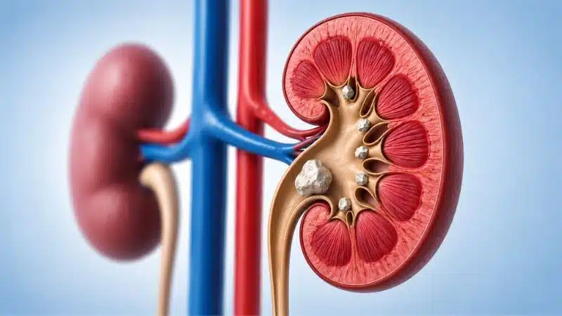
- Kidney stones or calcifications.
- Kidney cysts, which are fluid-filled sacs.
- Solid masses or growths in the kidney.
- Swelling of the kidney due to blockage, called hydronephrosis.
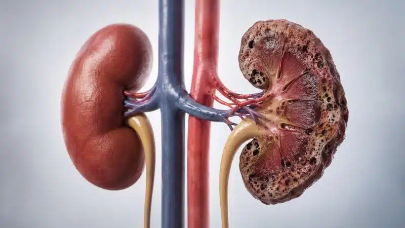
- Shrunken or small kidneys suggesting chronic disease.
- Congenital abnormalities such as extra kidney or malposition.
- Signs of infection or inflammation in some cases.
- Fluid collection around the kidney.
Ureter-related findings
- Dilatation of the ureter suggesting blockage.
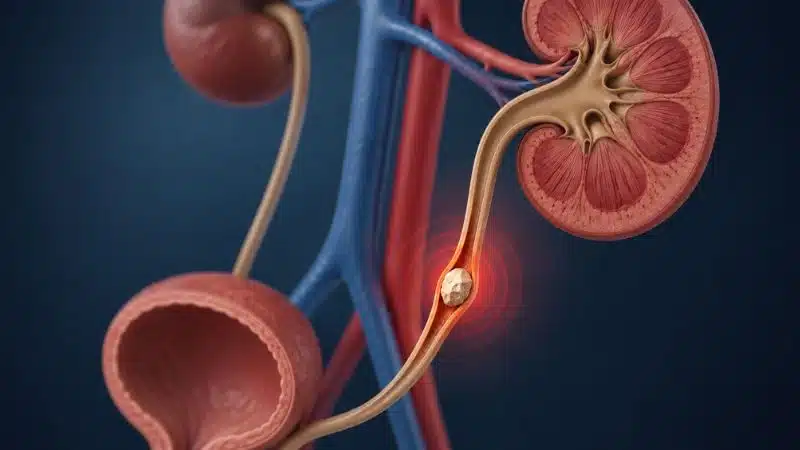
- Indirect signs of ureteric stones when the ureter is swollen.
- Compression of the ureter by nearby structures.
- Post-surgical changes in the ureter.
Bladder-related findings
- Bladder stones.
- Bladder wall thickening.
- Bladder masses or growths.
- Residual urine after voiding, suggesting incomplete emptying.
- Diverticula or outpouchings of the bladder wall.
- Signs of inflammation or infection.
- Abnormal bladder shape or size.
Other related findings
- Fluid in the abdomen near the urinary organs.
- Enlarged prostate in men causing bladder changes.
- Pregnancy-related changes affecting the urinary tract.
- Post-operative changes after kidney or bladder surgery.
Preparation for Ultrasound KUB
Proper preparation helps get clear images and accurate results. Instructions may vary slightly between centers, but some general points are commonly advised.
General preparation pointers
- Drink water before the test so the bladder is moderately full.
- A full bladder helps in better visualization of the bladder and lower ureters.
- Avoid emptying the bladder just before the scan unless instructed.
- Wear comfortable, loose-fitting clothing.
- Remove jewelry or metal objects from the abdomen area.
- Inform the staff if you are pregnant or have any medical conditions.
- Carry previous reports or scans if available for comparison.
Fasting requirements
- Some centers advise mild fasting for a few hours if other abdominal organs are also being checked.
- For a pure KUB scan, fasting is often not mandatory.
- Follow the specific instructions given by the radiology center.
Medicines and regular routine
- Continue regular medicines unless the doctor advises otherwise.
- Diabetic patients should follow individualized advice regarding food and medicines.
- If you have difficulty holding urine, inform the staff so they can adjust the timing.
How is Ultrasound KUB Performed?
The procedure is simple, painless, and usually completed within a short time.
Step-by-step procedure
- The patient is asked to lie on an examination table, usually on the back.
- A water-based gel is applied on the abdomen and lower belly area.
- The sonographer moves a handheld transducer over the skin.
- Images of the kidneys are taken from the back and sides.
- Images of the bladder are taken from the lower abdomen.
- The patient may be asked to change position for better views.
- The patient may be asked to empty the bladder and then scan may be repeated to check residual urine.
- The gel is wiped off after the scan.
- The images are reviewed by a radiologist.
Duration of the test
- The scan usually takes about fifteen to thirty minutes.
- It may take longer if the anatomy is complex or if additional views are needed.
- The patient can resume normal activity immediately after the test.
What the patient feels?
- The test is generally painless.
- There may be mild pressure from the transducer.
- A full bladder may cause some discomfort, but it is temporary.
- The gel may feel cool on the skin.
Benefits of Ultrasound KUB
Ultrasound KUB has several advantages that make it a preferred first-line test for urinary problems.
Key benefits
- No radiation exposure, making it safe for repeated use.
- Non-invasive and painless procedure.
- Can be done in children and pregnant women when needed.
- No need for contrast dye in most cases.
- Quick and widely available in most hospitals.
- Helps detect stones, cysts, blockages, and structural problems.
- Useful for follow-up of known kidney or bladder conditions.
- Cost-effective compared to some other imaging modalities.
- Real-time imaging allows dynamic assessment of the bladder.
Why it is often the first test?
- It gives a good overview of kidney size and structure.
- It can quickly identify hydronephrosis or large stones.
- It helps decide whether further tests like CT or MRI are needed.
- It is safe for screening and repeated monitoring.
Limitations of Ultrasound KUB
While Ultrasound KUB is very useful, it does have some limitations that patients should understand. In such cases, CT KUB, CT urography, MR urography, or other specialized tests may be advised.
Common limitations
- Small ureteric stones may not be seen directly.
- Obesity can reduce image quality in some cases.
- Gas in the intestines can block the view of deeper structures.
- Very small kidney stones may be missed.
- Detailed assessment of ureters is limited compared to CT.
- Some complex masses may need further imaging for characterization.
- Operator dependence means image quality can vary with expertise.
When additional tests may be needed
- If stone is strongly suspected but not seen on ultrasound.
- If a mass or growth needs detailed evaluation.
- If complex congenital anomalies are suspected.
- If detailed vascular assessment is required.
- If cancer staging or detailed anatomical mapping is needed.
Safety of Ultrasound KUB
Ultrasound KUB is considered very safe for most patients.
Safety pointers
- No ionizing radiation is used.
- No needles or injections are needed in standard scans.
- No known harmful effects from diagnostic ultrasound when used properly.
- Safe for children, elderly, and pregnant women when indicated.
- Can be repeated as needed for follow-up.
Special situations
- Pregnant women can safely undergo Ultrasound KUB when medically required.
- Children often tolerate the test well with proper explanation.
- Patients with pacemakers or implants can undergo ultrasound safely.
- Patients with kidney failure can have the test without contrast risk.
Who Should Get Ultrasound KUB?
Ultrasound KUB is recommended for many patients with urinary or abdominal symptoms.
Common indications
- Patients with flank or lower abdominal pain.
- Patients with blood in urine.
- Patients with recurrent urinary infections.
- Patients with suspected kidney stones.
- Patients with high blood pressure and suspected kidney cause.
- Patients with swelling in legs or face.
- Patients with abnormal kidney function tests.
- Patients with known kidney disease needing follow-up.
- Patients with bladder symptoms such as frequent urination or difficulty passing urine.
- Children with urinary tract infections or congenital anomalies.
- Pregnant women with urinary symptoms when advised by the doctor.
When it may not be the first choice?
- When detailed stone mapping is needed, CT KUB may be preferred.
- When cancer evaluation is needed, contrast-enhanced scans may be advised.
- When ureter detail is critical, specialized urography may be needed.
What Happens After Ultrasound KUB?
After the scan, the patient can usually resume normal routine immediately.
Post-test steps
- Wipe off the gel from the abdomen.
- Empty the bladder if it was kept full for the scan.
- Wait for the radiologist to prepare the report.
- Share the report with the treating doctor.
- Discuss further tests or treatment based on findings.
Report and interpretation
- The report describes kidney size, shape, and echotexture.
- It mentions any stones, cysts, masses, or hydronephrosis.
- It comments on bladder wall, capacity, and residual urine.
- It may suggest further tests if needed.
- The treating doctor correlates the report with symptoms and lab tests.
Ultrasound KUB in Special Groups
Ultrasound KUB is used across different age groups and conditions with some specific considerations.
In children
- Often the first imaging test for urinary infections or congenital issues.
- Helps detect structural abnormalities present from birth.
- Safe for repeated follow-up without radiation.
- May require parental support to keep the child calm.
In pregnant women
- Used when urinary symptoms need evaluation during pregnancy.
- Avoids radiation exposure to the mother and fetus.
- Helps assess kidney swelling or stones when suspected.
- Done only when clearly indicated by the doctor.
In elderly patients
- Useful for evaluating prostate-related bladder changes.
- Helps assess chronic kidney disease and structural changes.
- Safe even with multiple health conditions.
- Often part of routine work-up for urinary complaints.
In patients with chronic kidney disease
- Used to monitor kidney size and structure over time.
- Helps detect obstruction or worsening hydronephrosis.
- Safe for repeated scans during long-term follow-up.
- Guides timing of further interventions if needed.
Why Choose Rama Hospital for Ultrasound KUB?
Ultrasound KUB may seem like a simple test, but accurate imaging and correct interpretation are essential for proper diagnosis and treatment. A hospital with experienced radiologists, modern ultrasound machines, and coordinated urology and nephrology support can provide high-quality care.
- Experienced radiologists specializing in abdominal and urinary imaging.
- High-resolution ultrasound machines for better image quality.
- Proper protocol for KUB scanning and reporting.
- Coordination with urology and nephrology teams for further care.
- Quick report turnaround for timely treatment decisions.
Frequently Asked Questions (FAQs)
Is Ultrasound KUB painful?
No, Ultrasound KUB is not painful. It is a non-invasive test where a transducer is moved over the abdomen with some gel. The patient may feel mild pressure, but there is no cutting, needles, or radiation involved.
Do I need to fast for Ultrasound KUB?
Fasting is not always required for a pure KUB scan, but some centers advise mild fasting if other abdominal organs are also being checked. A full bladder is often recommended, so drinking water before the test is important.
How long does the test take?
Ultrasound KUB usually takes about fifteen to thirty minutes. It may take longer if the anatomy is complex or if additional views are needed. The patient can resume normal activity immediately after the test.
Can Ultrasound KUB detect all kidney stones?
Ultrasound KUB can detect many kidney stones, especially larger ones. However, small ureteric stones or very tiny stones may be missed. If stone is strongly suspected but not seen, CT KUB may be advised for better detection.
Is Ultrasound KUB safe in pregnancy?
Yes, Ultrasound KUB is considered safe in pregnancy when medically indicated because it does not use radiation. It is often preferred over CT scans for evaluating urinary symptoms in pregnant women under doctor’s guidance.
Do I need to keep my bladder full for the scan?
Yes, a moderately full bladder helps in better visualization of the bladder and lower ureters. Patients are usually advised to drink water before the test and avoid emptying the bladder just before the scan unless instructed otherwise.
What happens if the report is abnormal?
If the report is abnormal, the treating doctor will correlate it with symptoms and other tests. Further evaluation, treatment, or referral to a urologist or nephrologist may be advised depending on the findings.
Conclusion
Ultrasound KUB is a safe, non-invasive, and widely used imaging test for evaluating the kidneys, ureters, and bladder. It helps detect stones, cysts, blockages, structural abnormalities, and other urinary problems without radiation or contrast dye in most cases. It is often the first test advised for patients with urinary symptoms, abdominal pain, or suspected kidney disease.
For patients with flank pain, urinary infections, blood in urine, or abnormal kidney tests, Ultrasound KUB provides valuable information that guides further management. With proper preparation, expert scanning, and accurate reporting, it plays a key role in urology and nephrology care.
Rama Hospital can provide high-quality Ultrasound KUB imaging with experienced radiologists, modern equipment, and coordinated support from urology and nephrology teams for comprehensive urinary care.