The heart is a complex organ that works continuously to pump blood to every part of the body. To understand how well it is functioning, doctors need detailed and accurate images of its structure and movement. One of the most advanced tools available today for this purpose is 3D echocardiography. Unlike a regular 2D ultrasound of the heart, 3D echocardiography creates a three-dimensional view of the heart’s chambers, valves, and blood flow. This gives doctors a much clearer and more complete picture of how the heart is working.
3D echocardiography is a non-invasive test, which means it does not require surgery, cuts, or radiation. It uses sound waves to create moving images of the heart in real time. Because of its high level of detail, it is especially useful in evaluating heart valves, congenital heart defects, complex heart conditions, and patients undergoing certain procedures. It helps doctors make better decisions about diagnosis, treatment, and follow-up.
What is 3D Echocardiography?
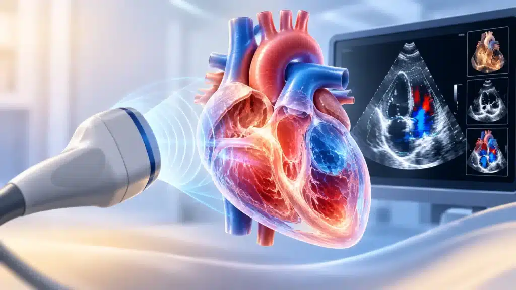
3D echocardiography is an advanced form of ultrasound imaging that creates a three-dimensional view of the heart. It uses high-frequency sound waves that bounce off the heart structures and return to a special probe. A computer then converts these echoes into detailed moving images.
Unlike a standard 2D echocardiogram, which shows flat, two-dimensional slices of the heart, 3D echo allows doctors to see the heart from multiple angles and depths. This makes it easier to understand the true shape, size, and movement of heart chambers and valves. It is especially helpful for complex cases where a 2D view may not give enough information.
The test is safe, painless, and does not use radiation. It can be done as part of a routine heart check-up or as a detailed evaluation for specific heart problems. In many hospitals, 3D echocardiography is used alongside other cardiac tests to give a complete picture of heart health.
How Does 3D Echocardiography Work?
The test works on the same basic principle as ultrasound used in pregnancy or abdominal scans, but it is adapted for the heart.
- A small handheld probe called a transducer is placed on the chest.
- The probe sends high-frequency sound waves into the body.
- These waves bounce off the heart structures and return as echoes.
- A computer processes the echoes to create detailed images.
- In 3D mode, multiple image planes are combined to form a three-dimensional view.
- Doctors can rotate, slice, and examine the heart from different angles on the screen.
Types of 3D echo views
- Real-time 3D imaging, which shows the heart moving in real time.
- Full-volume 3D imaging, which captures a complete 3D dataset of the heart.
- 3D transesophageal echo, where the probe is placed in the food pipe for closer views of the heart.
Who Needs 3D Echocardiography?
Not every patient needs a 3D echo. It is usually recommended when more detail is needed than a standard 2D echo can provide.
Common reasons for the test
- Evaluation of heart valve disease.
- Assessment before or after valve repair or replacement.
- Evaluation of congenital heart defects.
- Assessment of complex heart anatomy.
- Planning for cardiac surgery or catheter-based procedures.
- Evaluation of heart function in difficult cases.
- Assessment of masses, clots, or abnormal structures inside the heart.
- Monitoring of known heart conditions over time.
Patients who may benefit
- People with suspected or known valve disease.
- Patients with congenital heart disease.
- People being evaluated for heart surgery.
- Patients with unclear findings on 2D echo.
- Those with complex heart rhythm problems.
- Individuals with a history of heart infection affecting valves.
- Patients undergoing structural heart procedures.
What Can 3D Echocardiography Detect?
Because of its detailed imaging, 3D echo can identify many heart problems more clearly than standard tests.
Heart valve problems
- Narrowing of valves such as aortic or mitral stenosis.
- Leaking valves such as mitral or aortic regurgitation.
- Structural damage to valve leaflets.
- Prosthetic valve function and complications.
- Valve infection or vegetations.
Heart chamber and function issues
- Enlarged heart chambers.
- Reduced pumping function.
- Abnormal wall motion.
- Heart failure assessment.
- Assessment of left ventricle size and function.
Congenital and structural problems
- Holes in the heart such as atrial or ventricular septal defects.
- Abnormal connections between chambers or vessels.
- Complex congenital anatomy.
- Structural abnormalities seen from birth.
Other findings
- Blood clots inside the heart.
- Tumors or masses.
- Fluid around the heart.
- Complications after heart procedures.
- Assessment before structural heart interventions.
Benefits of 3D Echocardiography
3D echo offers several advantages over standard 2D imaging.
Key benefits
- More detailed and accurate view of heart structures.
- Better assessment of valve shape and function.
- Improved detection of complex defects.
- Better planning for surgery or procedures.
- Non-invasive with no radiation.
- Real-time imaging of heart movement.
- Useful for both diagnosis and follow-up.
- Helps reduce uncertainty in difficult cases.
Why accuracy matters
In heart care, small details can make a big difference. For example, the exact shape of a leaking valve or the size of a hole in the heart can change the treatment plan. 3D echo gives doctors the detail they need to choose the best approach for each patient.
Types of 3D Echocardiography
There are different ways this test can be done depending on the patient and the question being answered.
1. Transthoracic 3D echocardiography
This is the most common type, where the probe is placed on the chest wall.
- Non-invasive and painless.
- Suitable for most patients.
- Used for routine and detailed heart evaluation.
- May have limited views in some patients due to body habitus or lung disease.
2. Transesophageal 3D echocardiography
In this type, a special probe is passed into the food pipe to get closer views of the heart.
- Provides very detailed images, especially of valves.
- Useful for complex valve disease and congenital defects.
- Often used before or during heart procedures.
- Requires fasting and mild sedation in many cases.
3. 3D echo during procedures
Sometimes 3D echo is used during heart procedures to guide the doctor in real time.
- Helps guide catheter-based valve procedures.
- Used during certain structural heart interventions.
- Improves safety and accuracy.
- Allows immediate assessment of results.
Preparation for the Test
Preparation depends on the type of 3D echo being done.
For transthoracic 3D echo
- No special preparation is usually needed.
- Wear comfortable clothing that allows easy access to the chest.
- Continue regular medicines unless advised otherwise.
- Inform the team about any heart devices or allergies.
For transesophageal 3D echo
- Fasting for several hours before the test is usually required.
- Inform the team about allergies, swallowing problems, or esophageal issues.
- Arrange for someone to accompany you if sedation is used.
- Follow specific instructions given by the hospital.
What Happens During the Test?
The experience is different for transthoracic and transesophageal 3D echo.
During transthoracic 3D echo
- You will lie on an examination table.
- Gel is applied to the chest to improve sound wave transmission.
- The probe is moved over different areas of the chest.
- You may be asked to breathe in a certain way or hold your breath briefly.
- The test usually takes 20 to 45 minutes.
- You can return to normal activities immediately after.
During transesophageal 3D echo
- A throat spray or mild sedation may be given.
- A thin, flexible probe is passed into the food pipe.
- Images of the heart are taken from close range.
- The test usually takes 30 to 60 minutes.
- You may need to rest and avoid eating for a short time after.
Risks and Safety
3D echocardiography is very safe, especially the transthoracic type.
Safety of transthoracic 3D echo
- No radiation exposure.
- No needles or cuts.
- No known serious risks.
- Safe for repeated use and follow-up.
Safety of transesophageal 3D echo
- Small risk of throat discomfort or irritation.
- Rare risk of injury to the food pipe.
- Risks related to sedation in some patients.
- Overall very safe when done by trained teams.
Recovery After the Test
Recovery depends on the type of test.
After transthoracic 3D echo
- No recovery time needed.
- Can resume normal activities immediately.
- Gel on chest can be wiped off easily.
After transesophageal 3D echo
- May feel throat discomfort or mild soreness.
- Avoid eating or drinking until advised.
- Rest for a short period if sedation was used.
- Arrange for someone to take you home if sedated.
How 3D Echo Compares with Other Heart Tests
Different heart tests give different types of information.
3D echo vs 2D echo
- 3D echo gives a more complete, three-dimensional view.
- Better for complex valve and structural problems.
- 2D echo is still useful for routine assessment.
3D echo vs ECG
- ECG records electrical activity of the heart.
- 3D echo shows structure and movement.
- Both tests give different but complementary information.
3D echo vs cardiac MRI or CT
- MRI and CT give detailed images but may involve radiation or longer scan times.
- 3D echo is real-time, radiation-free, and often easier to repeat.
- Choice depends on the clinical question and patient condition.
When is 3D Echo Recommended?
Doctors may recommend 3D echo in specific situations.
Common recommendations
- Before valve repair or replacement surgery.
- During evaluation of complex congenital heart disease.
- When 2D echo findings are unclear.
- For detailed assessment of prosthetic valves.
- During structural heart procedures.
- For follow-up of known heart conditions.
Why Choose Rama Hospital for 3D Echocardiography?
Advanced heart imaging requires skilled doctors, modern equipment, and a coordinated care team. A hospital with experienced cardiologists, cardiac imaging specialists, and support staff can make a major difference in accuracy and patient experience.
Why hospital care matters
- Access to advanced 3D echo technology.
- Experienced cardiologists and imaging specialists.
- Detailed and accurate reporting.
- Coordination with surgery or interventional teams when needed.
- Comprehensive cardiac evaluation under one roof.
Trust and authority points
- Modern cardiac imaging facilities.
- Experienced heart care team.
- Clear explanation of results and next steps.
- Patient-centered approach.
- Support for both diagnosis and treatment planning.
Frequently Asked Questions (FAQs)
Is a 3D echo different from a normal echo?
Yes, a normal echo is usually 2D and shows flat images of the heart. 3D echo adds depth and allows doctors to view the heart from multiple angles. This is especially useful for complex valve problems or structural heart defects.
Is 3D echocardiography painful?
No, the transthoracic 3D echo is painless and non-invasive. The transesophageal type may cause mild throat discomfort, but sedation and numbing spray are used to keep the patient comfortable.
How long does the test take?
A transthoracic 3D echo usually takes 20 to 45 minutes. A transesophageal 3D echo may take 30 to 60 minutes, including preparation and recovery time.
Do I need to fast before the test?
For a transthoracic 3D echo, fasting is usually not required. For a transesophageal 3D echo, fasting for several hours is usually needed. Always follow the specific instructions given by the hospital.
Is there any radiation in 3D echo?
No, 3D echocardiography uses sound waves, not X-rays. This means there is no radiation exposure, making it safe for repeated use and follow-up.
Can 3D echo detect all heart problems?
3D echo is very useful for valves, chambers, and structural problems, but it may not detect every issue. Doctors may combine it with ECG, stress tests, CT, or MRI depending on the clinical question.
When will I get the results?
In many cases, preliminary findings are discussed soon after the test. A detailed report may take a little longer depending on the complexity and hospital workflow. Your doctor will explain the results and next steps.
Conclusion
3D echocardiography is an advanced, safe, and highly detailed heart imaging test that gives doctors a much clearer view of the heart’s structure and function. It is especially valuable for evaluating heart valves, congenital defects, and complex cardiac conditions. Because it is non-invasive and radiation-free, it can be used safely for both diagnosis and follow-up.
For patients with known or suspected heart problems, 3D echo can provide the detailed information needed to plan the right treatment. Whether it is used for routine assessment, pre-surgical planning, or guidance during procedures, this test plays an important role in modern cardiac care.
Rama Hospital can provide advanced 3D echocardiography along with expert cardiac evaluation, clear reporting, and coordinated care for patients who need detailed heart assessment.