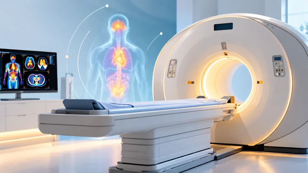
A PET scan, or Positron Emission Tomography scan, is an advanced imaging test that helps doctors see how organs and tissues are working inside the body. Unlike a regular X-ray or CT scan that mainly shows structure, a PET scan shows function and activity at the cellular level. This makes it very useful in detecting diseases early, understanding how active a condition is, and guiding treatment decisions.
PET scans are commonly used in cancer care, heart disease, brain disorders, and infection or inflammation-related conditions. In many cases, a PET scan is combined with a CT scan or MRI scan to give doctors both detailed structure and functional information in one test. This combination helps in accurate diagnosis, staging of disease, treatment planning, and follow-up.
What is a PET Scan?
A PET scan is a type of nuclear medicine imaging test. It uses a small amount of radioactive tracer, usually injected into a vein, to show how tissues and organs are functioning. The tracer collects in areas of higher activity, such as cancer cells, inflamed tissue, or active brain areas. A special camera then detects the signals from the tracer and creates detailed images.
Because PET scan shows activity rather than just structure, it can detect changes in the body before they become visible on other imaging tests. This is why it is often used in early detection, staging, and monitoring of disease.
In many hospitals, PET scan is combined with CT scan or MRI scan. This gives doctors both functional information from the PET part and detailed anatomical information from the CT or MRI part. The combined test is called PET CT or PET MRI.
Why is a PET Scan Done?
PET scans are done for many reasons depending on the suspected condition and the information the doctor needs.
Common reasons for PET scan
- Detect cancer or abnormal growths.
- Determine the stage of cancer.
- Check whether cancer has spread to other parts of the body.
- Evaluate how well cancer treatment is working.
- Detect recurrence of cancer after treatment.
- Assess heart function and blood flow.
- Evaluate brain activity in certain neurological conditions.
- Detect infection or inflammation in the body.
- Guide biopsy or treatment planning.
- Monitor response to therapy over time.
Why PET scan is useful
- Shows activity at the cellular level.
- Can detect disease earlier than some other tests.
- Helps in accurate staging and treatment planning.
- Combines functional and structural information when paired with CT or MRI.
- Reduces the need for multiple separate tests in some cases.
Types of PET Scan
PET scans can be classified based on the tracer used and the area of the body being studied.
Based on tracer used
- FDG PET scan, which uses a glucose-based tracer and is most common in cancer and inflammation imaging.
- Cardiac PET scan, which uses tracers to assess blood flow and heart function.
- Brain PET scan, which uses tracers to study brain activity and certain neurological conditions.
- Bone PET scan, which uses tracers to evaluate bone activity or infection.
Based on combination with other imaging
- PET CT scan, which combines PET with CT for detailed structure and function.
- PET MRI scan, which combines PET with MRI for advanced soft tissue detail.
- Standalone PET scan, which uses PET imaging alone.
Based on purpose
- Oncology PET scan, used mainly for cancer detection, staging, and follow-up.
- Cardiac PET scan, used for heart disease evaluation.
- Neurology PET scan, used for brain and nerve-related conditions.
- Infection or inflammation PET scan, used to detect hidden infection or inflammatory disease.
Conditions Commonly Evaluated with PET Scan
PET scans are used across many medical specialties.
Cancer-related conditions
- Lung cancer.
- Breast cancer.
- Lymphoma.
- Colorectal cancer.
- Head and neck cancers.
- Melanoma.
- Esophageal cancer.
- Pancreatic cancer.
- Recurrent or metastatic cancer.
Heart-related conditions
- Coronary artery disease.
- Heart muscle viability assessment.
- Evaluation after heart attack.
- Assessment of blood flow to the heart.
Brain and neurological conditions
- Epilepsy.
- Alzheimer’s disease and other dementias.
- Brain tumors.
- Parkinson’s disease and movement disorders.
- Evaluation of brain function in selected cases.
Infection and inflammation
- Hidden infection or abscess.
- Inflammatory diseases.
- Evaluation of fever of unknown origin.
- Assessment of inflammation in blood vessels or organs.
How to Prepare for a PET Scan
Preparation depends on the type of PET scan and the area being studied. Following instructions carefully helps ensure accurate results.
General preparation points
- Follow fasting instructions if advised, usually 4 to 6 hours before the scan.
- Avoid strenuous exercise for 24 hours before the scan.
- Inform the doctor about all medicines, supplements, and allergies.
- Inform the team if you are pregnant or breastfeeding.
- Remove metal objects such as jewelry, watches, or dentures before the scan.
- Wear comfortable, loose clothing without metal parts.
Preparation for cancer PET scan
- Fasting is usually required to keep blood sugar low for better tracer uptake.
- Avoid sugary drinks and high-carbohydrate meals before fasting.
- Diabetic patients may need special instructions for insulin or medicines.
- Drink water as advised to stay hydrated.
Preparation for cardiac PET scan
- Avoid caffeine for 24 hours before the scan.
- Follow instructions about food and medicines.
- Inform the team about heart medicines or blood thinners.
Preparation for brain PET scan
- Follow fasting instructions if advised.
- Avoid caffeine and certain medicines if instructed.
- Inform the team about neurological conditions or medicines.
What Happens During a PET Scan?
Knowing what to expect can reduce anxiety and help the scan go smoothly.
Before the scan
- The patient changes into a hospital gown if needed.
- A small IV line may be placed in the arm.
- The radioactive tracer is injected through the IV.
- The patient rests quietly for 30 to 60 minutes while the tracer circulates.
- Blood sugar may be checked in some cases.
During the scan
- The patient lies on a padded table that slides into the PET scanner.
- The scanner is a large, ring-shaped machine.
- The patient must remain still during the scan.
- The scan usually takes 20 to 45 minutes depending on the area studied.
- The technologist may give instructions through an intercom.
After the scan
- The patient may be asked to wait briefly to ensure there are no reactions.
- Drinking water helps flush the tracer from the body.
- Normal activities can usually be resumed unless advised otherwise.
- Results are reviewed by a radiologist or nuclear medicine specialist.
Benefits of PET Scan
PET scan offers several advantages over other imaging tests in certain situations.
Key benefits
- Detects disease at the cellular level before structural changes appear.
- Helps in early diagnosis of cancer and other conditions.
- Provides accurate staging of cancer.
- Helps in treatment planning and monitoring.
- Combines functional and anatomical information with CT or MRI.
- Reduces the need for multiple tests in some cases.
- Helps assess treatment response and detect recurrence.
- Guides biopsy or surgery in selected cases.
Benefits in cancer care
- Detects active cancer cells.
- Shows spread of cancer to lymph nodes or other organs.
- Helps decide between surgery, chemotherapy, radiation, or other treatments.
- Monitors response to therapy.
- Detects recurrence early.
Benefits in heart and brain care
- Assesses blood flow and heart muscle viability.
- Helps in planning treatment for coronary artery disease.
- Evaluates brain activity in epilepsy or dementia.
- Helps in diagnosis and monitoring of neurological conditions.
Risks and Safety of PET Scan
PET scan is generally safe, but it involves exposure to a small amount of radiation. The benefits usually outweigh the risks when the test is medically indicated.
Radiation exposure
- The tracer contains a small amount of radioactive material.
- Radiation exposure is low and similar to other diagnostic imaging tests.
- The tracer breaks down and leaves the body through urine within hours.
Allergic reactions
- Rare, but possible.
- Inform the team about any previous reactions to contrast dye or tracers.
- Emergency support is available if needed.
Pregnancy and breastfeeding
- PET scan is generally avoided in pregnancy unless absolutely necessary.
- Breastfeeding mothers may need to pause feeding for a short time after the scan.
- Inform the team if pregnant or breastfeeding before the scan.
Kidney or other health concerns
- Inform the doctor if you have kidney disease, diabetes, or other chronic conditions.
- Special instructions may be needed for diabetic patients.
- Hydration and medicine adjustments may be advised.
What to Expect After a PET Scan
Most patients can return to normal activities soon after the scan.
After the scan
- Drink plenty of water to help flush the tracer.
- Resume normal diet unless advised otherwise.
- Avoid close contact with pregnant women or young children for a few hours if advised.
- Follow any specific instructions given by the team.
- Results are usually discussed at a follow-up appointment.
Understanding results
- Areas of high tracer uptake may indicate active disease, inflammation, or cancer.
- The radiologist compares PET images with CT or MRI images if combined.
- Results help guide diagnosis, staging, and treatment decisions.
PET Scan vs Other Imaging Tests
PET scan is different from other imaging tests in what it shows.
PET scan vs CT scan
- CT shows detailed structure and anatomy.
- PET shows function and activity.
- PET CT combines both for more complete information.
PET scan vs MRI scan
- MRI shows detailed soft tissue images.
- PET shows metabolic activity.
- PET MRI combines both for advanced evaluation.
PET scan vs X-ray
- X-ray is quick and shows bones and some organs.
- PET shows cellular activity and is more sensitive for certain diseases.
- PET is more detailed but takes longer and involves tracer injection.
When is PET Scan Recommended?
PET scan is recommended when the doctor needs detailed functional information that other tests cannot provide.
Common situations
- Suspected cancer or unexplained mass.
- Staging of known cancer.
- Evaluation of treatment response.
- Detection of cancer recurrence.
- Assessment of heart blood flow.
- Evaluation of brain disorders.
- Detection of hidden infection or inflammation.
- Guidance for biopsy or surgery.
Why timing matters
- Early PET scan can detect disease before it spreads.
- Follow-up PET scan can assess treatment response.
- Timing depends on the condition and treatment plan.
Why Choose Rama Hospital for PET Scan?
PET scan requires advanced technology, trained nuclear medicine specialists, and careful safety protocols. A hospital with experienced radiology and nuclear medicine teams can ensure accurate results and safe care.
Why hospital care matters
- Advanced PET CT and PET MRI facilities.
- Experienced radiologists and nuclear medicine specialists.
- Safe tracer handling and radiation safety protocols.
- Comfortable and patient-friendly environment.
- Coordination with oncology, cardiology, neurology, and other departments.
- Clear communication of results and next steps.
Trust and authority points
- Modern imaging technology.
- Trained and certified staff.
- Strict safety and quality protocols.
- Patient education and support.
- Comprehensive care from diagnosis to treatment planning.
Frequently Asked Questions (FAQs)
1. What is a PET scan?
A PET scan is an imaging test that shows how organs and tissues are functioning. It uses a small amount of radioactive tracer to detect activity at the cellular level. It is often combined with CT or MRI for detailed information.
2. Why is a PET scan done?
PET scan is done to detect cancer, assess its spread, monitor treatment response, evaluate heart or brain conditions, and detect infection or inflammation. It gives functional information that other tests may not show.
3. How should I prepare for a PET scan?
Preparation may include fasting for 4 to 6 hours, avoiding strenuous exercise, and informing the team about medicines and allergies. Diabetic patients may need special instructions. Follow the specific advice given by the hospital.
4. Is a PET scan safe?
Yes, PET scan is generally safe. It involves a small amount of radiation from the tracer, which leaves the body within hours. The benefits usually outweigh the risks when the test is medically needed.
5. How long does a PET scan take?
The scan itself usually takes 20 to 45 minutes. Including tracer injection and waiting time, the whole process may take 2 to 3 hours. The exact time depends on the type of scan and area studied.
6. Can I eat before a PET scan?
For many PET scans, especially cancer scans, fasting is required. Avoid sugary drinks and high-carbohydrate meals before fasting. Follow the specific instructions given by the hospital.
7. Is PET scan painful?
The scan itself is painless. The only discomfort may be from the IV needle when the tracer is injected. The patient must lie still during the scan, but there is no pain from the imaging itself.
8. Can pregnant women have a PET scan?
PET scan is generally avoided in pregnancy unless absolutely necessary. If a scan is needed, the team will take special precautions. Always inform the staff if you are pregnant or breastfeeding.
9. How soon will I get PET scan results?
Results are usually reviewed by a radiologist or nuclear medicine specialist and discussed at a follow-up appointment. In urgent cases, results may be available sooner. The timing depends on the hospital and the type of scan.
10. What is the difference between PET CT and PET MRI?
PET CT combines PET with CT scan for detailed structure and function. PET MRI combines PET with MRI for advanced soft tissue detail. The choice depends on the condition being evaluated and the doctor’s recommendation.
Conclusion
PET scan is an advanced imaging test that gives doctors detailed information about how organs and tissues are functioning. It is especially useful in cancer care, heart disease, brain disorders, and infection or inflammation-related conditions. By showing activity at the cellular level, PET scan can detect disease early, guide treatment, and monitor response over time.
The most important steps are proper preparation, following medical advice, and understanding what the scan involves. For patients with suspected cancer, heart disease, neurological conditions, or unexplained inflammation, a PET scan can make a major difference in diagnosis and treatment planning. With the right care and advanced imaging, patients can get accurate answers and better health outcomes.
Rama Hospital can help evaluate the need for PET scan, provide advanced imaging facilities, and guide patients through the process safely and comfortably.