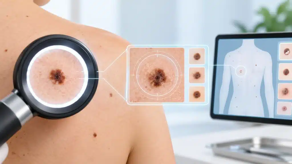
Digital mole mapping is a modern, non-invasive method used to monitor moles and other pigmented skin lesions over time. It involves taking high-resolution, standardized photographs of the skin surface and storing them digitally for future comparison. This allows dermatologists to detect even subtle changes in moles that may not be visible to the naked eye during a routine examination.
The main purpose of digital mole mapping is early detection of skin cancer, especially melanoma, which is one of the most serious types of skin cancer. When detected early, skin cancer is highly treatable. Digital mapping provides a reliable baseline record of all moles on the body, making it easier to identify new lesions or changes in existing ones during follow-up visits.
What is Digital Mole Mapping?
Digital mole mapping, also known as total body photography or digital dermoscopy, is a specialized imaging technique used to document and monitor moles and other skin spots. It uses high-quality cameras and sometimes dermoscopic lenses to capture detailed images of the skin surface.
The process creates a visual record of all moles on the body at a specific point in time. These images are stored securely and can be compared with future images to check for changes. Even small changes in size, shape, color, or structure can be detected more accurately with digital comparison than with memory or hand-drawn maps.
Digital mole mapping does not diagnose skin cancer on its own. Instead, it supports the dermatologist in deciding which moles need closer examination, biopsy, or removal. It is especially useful for people with many moles, atypical moles, or higher risk of skin cancer.
Why is Digital Mole Mapping Done?
The main reason for digital mole mapping is early detection of skin cancer. Melanoma and other skin cancers often start as changes in existing moles or as new pigmented lesions. Detecting these changes early improves treatment outcomes significantly.
Key reasons for digital mole mapping
- To create a baseline record of all moles on the body.
- To monitor moles over time for changes.
- To detect new moles that appear between visits.
- To identify subtle changes that may suggest early skin cancer.
- To reduce unnecessary biopsies of stable, benign moles.
- To provide peace of mind for high-risk patients.
- To support regular skin cancer screening in a structured way.
Why early detection matters
Skin cancer, especially melanoma, can spread if not detected early. Early-stage melanoma is often curable with simple surgical removal. Advanced melanoma may require more complex treatment and has a higher risk of complications. Digital mole mapping helps catch suspicious changes at the earliest possible stage.
Who Should Consider Digital Mole Mapping?
Not everyone needs digital mole mapping, but it is particularly useful for people at higher risk of skin cancer or those with many moles.
Higher-risk groups
- People with a large number of moles, especially more than 50.
- People with atypical or irregular moles.
- Individuals with a personal history of melanoma or other skin cancer.
- People with a family history of melanoma.
- Individuals with very fair skin, light eyes, or red or blond hair.
- People who have had severe sunburns, especially in childhood.
- Those with significant sun exposure due to work or lifestyle.
- Individuals with a history of indoor tanning or UV bed use.
- People with weakened immune systems.
- Patients with genetic conditions that increase skin cancer risk.
Who may benefit even without high risk
- People who find it difficult to monitor their own skin.
- Individuals who want a structured skin health record.
- Patients who feel anxious about moles and want regular monitoring.
- People who have noticed new or changing moles and want careful follow-up.
How Does Digital Mole Mapping Work?
The procedure is simple, painless, and non-invasive. It is usually done in a dermatology clinic or hospital setting by trained staff.
Step-by-step procedure
- The patient is asked to remove clothing and wear a gown or shorts for the examination.
- The skin is examined to identify all moles and pigmented lesions.
- Standardized photographs are taken of the entire body or specific areas.
- High-resolution images are captured using specialized cameras and lighting.
- In some cases, close-up dermoscopic images of specific moles are also taken.
- The images are stored securely in a digital system linked to the patient’s record.
- The dermatologist reviews the images and notes any moles that need closer attention.
Duration and comfort
- The procedure usually takes 20 to 40 minutes depending on the number of moles and areas covered.
- It is painless and does not involve radiation or needles.
- Patients may feel slightly exposed, but privacy and comfort are maintained throughout.
- No special preparation is needed, but patients should avoid heavy lotions or makeup on the day of mapping.
What Happens During Follow-Up?
Digital mole mapping is most useful when repeated over time. Follow-up visits allow comparison between current and previous images.
Follow-up process
- The patient returns for a repeat mapping session, usually after 6 to 12 months, or as advised.
- New images are taken using the same standardized method.
- The dermatologist compares new images with baseline and previous images.
- Any new moles or changes in existing moles are identified.
- Moles that show suspicious changes may be examined more closely with dermoscopy.
- If needed, a biopsy or removal may be recommended for further testing.
Typical follow-up intervals
- Every 6 months for high-risk patients.
- Every 12 months for moderate-risk patients.
- As needed for patients with specific concerning moles.
- More frequently if a suspicious lesion is being monitored closely.
Benefits of Digital Mole Mapping
Digital mole mapping offers several advantages over routine visual examination alone.
Key benefits
- Early detection of skin cancer and precancerous changes.
- Accurate comparison of moles over time.
- Detection of new moles that may not be noticed by the patient.
- Reduced anxiety for patients worried about moles.
- Better documentation for medical records.
- Support for more informed decisions about biopsy or removal.
- Non-invasive and painless procedure.
- Useful for patients with many moles that are hard to track manually.
Why it is more effective than self-checks
Many people find it difficult to check their own back, scalp, or other hard-to-see areas. Even dermatologists may find it challenging to remember the exact appearance of every mole during a routine visit. Digital mapping provides an objective record that makes change detection more reliable.
Limitations of Digital Mole Mapping
While digital mole mapping is a powerful tool, it has some limitations that patients should understand.
Important limitations
- It does not diagnose skin cancer on its own.
- It cannot detect skin cancers that do not arise from moles.
- It requires regular follow-up to be effective.
- It may not be necessary for low-risk individuals with few moles.
- It does not replace the need for clinical examination and dermoscopy.
- It may not be available in all clinics or hospitals.
- It requires trained staff and proper equipment for best results.
Why clinical judgment still matters
Digital images support but do not replace the dermatologist’s expertise. A skilled dermatologist uses mapping images along with physical examination, dermoscopy, and patient history to make the best decisions about further testing or treatment.
Role in Skin Cancer Screening
Digital mole mapping is part of a broader skin cancer screening strategy. It works best when combined with regular skin checks, sun protection, and patient awareness.
Components of skin cancer screening
- Regular self-examination of the skin.
- Annual or periodic dermatologist visits for full-body skin checks.
- Digital mole mapping for high-risk or mole-prone individuals.
- Dermoscopy for detailed examination of suspicious moles.
- Biopsy or removal of lesions that appear suspicious.
- Education on sun protection and early warning signs.
How mapping fits into screening
Digital mole mapping provides a detailed baseline that makes future screening more accurate. It helps the dermatologist focus on moles that have changed rather than examining every mole in detail at every visit. This makes screening more efficient and targeted.
Preparation for Digital Mole Mapping
Preparation is simple, but a few steps can make the process smoother.
Before the appointment
- Avoid applying heavy lotions, oils, or makeup on the day of mapping.
- Remove nail polish if possible, as moles on fingers and toes may be photographed.
- Wear comfortable clothing that is easy to remove.
- Bring a list of any moles you are particularly concerned about.
- Inform the clinic if you have any mobility issues or need privacy accommodations.
What to expect on the day
- You will be asked to change into a gown or shorts.
- A trained technician or dermatologist will take the photographs.
- The process is systematic and covers all body areas.
- You can ask questions at any time during the procedure.
- The dermatologist will review the images and discuss any findings.
After the Procedure
There is no recovery time needed after digital mole mapping. Patients can resume normal activities immediately.
Post-procedure guidance
- Continue regular self-examination of your skin between visits.
- Follow the recommended follow-up schedule for repeat mapping.
- Report any new or changing moles to your dermatologist promptly.
- Practice sun protection to reduce future skin damage.
- Keep your contact information updated so the clinic can reach you if needed.
When to contact the clinic sooner
- If you notice a new mole that looks different from others.
- If an existing mole changes in size, shape, or color.
- If a mole starts bleeding, itching, or becoming painful.
- If you develop a non-healing spot or sore on the skin.
- If you have any concerns between scheduled visits.
Who Performs Digital Mole Mapping?
Digital mole mapping should be performed by trained healthcare professionals in a clinical setting.
Qualified providers
- Dermatologists with training in skin cancer screening.
- Trained dermatology nurses or technicians under medical supervision.
- Clinics or hospitals with proper imaging equipment and software.
- Healthcare settings that maintain patient privacy and data security.
Why professional setting matters
Proper lighting, camera quality, and standardized positioning are essential for accurate mapping. Professional settings also ensure that images are stored securely and linked to your medical record for future comparison.
Cost and Availability
Digital mole mapping may not be available in all clinics, and costs can vary depending on the facility and technology used.
Factors affecting cost
- Type of equipment used (standard photography vs. advanced dermoscopic mapping).
- Extent of mapping (full body vs. specific areas).
- Whether dermoscopic close-ups are included.
- Follow-up visits and comparison services.
- Hospital or clinic pricing policies.
Insurance and coverage
- Some insurance plans may cover mole mapping if it is part of skin cancer screening for high-risk patients.
- Many patients pay out-of-pocket for mapping as a preventive service.
- It is best to check with your insurance provider and the hospital beforehand.
Sun Protection and Skin Health
Digital mole mapping is most effective when combined with good sun protection and skin care habits.
Sun protection tips
- Use broad-spectrum sunscreen with SPF 30 or higher daily.
- Reapply sunscreen every 2 to 3 hours when outdoors.
- Wear protective clothing, hats, and sunglasses.
- Seek shade during peak sun hours, usually between 10 AM and 4 PM.
- Avoid indoor tanning or UV beds completely.
- Check your skin regularly for new or changing spots.
Why sun protection matters
UV exposure is the most significant preventable risk factor for skin cancer. Protecting your skin reduces the formation of new moles, prevents DNA damage, and lowers the risk of melanoma and other skin cancers over time.
Frequently Asked Questions (FAQs)
Is digital mole mapping painful?
No, digital mole mapping is completely painless. It involves taking photographs of the skin using specialized cameras. There are no needles, no radiation, and no discomfort during the procedure.
Who should get digital mole mapping?
It is most useful for people with many moles, atypical moles, a personal or family history of melanoma, fair skin, or significant sun exposure. It can also benefit anyone who wants structured skin monitoring and early cancer detection.
How often should I repeat mole mapping?
Most high-risk patients are advised to repeat mapping every 6 to 12 months. The exact interval depends on your risk level, number of moles, and dermatologist’s recommendation. Some patients may need more frequent monitoring.
Can digital mole mapping detect all skin cancers?
No, it cannot detect all skin cancers. It is most effective for detecting changes in moles and pigmented lesions. Some skin cancers arise in normal-looking skin and may not be captured by mapping alone, which is why regular skin checks are still important.
Does digital mole mapping replace biopsy?
No, digital mole mapping does not replace biopsy. It helps identify which moles need closer examination. If a mole looks suspicious, a biopsy or removal may still be needed to confirm whether it is cancerous.
Is digital mole mapping safe?
Yes, it is very safe. It uses visible light photography and does not involve radiation, needles, or invasive procedures. It is suitable for most patients, including children and pregnant women, when recommended by a dermatologist.
What happens if a mole changes on mapping?
If a mole shows changes, the dermatologist will examine it more closely, often with dermoscopy. Depending on the findings, a biopsy or removal may be recommended to rule out skin cancer. Early action improves treatment outcomes.
Conclusion
Digital mole mapping is a valuable, non-invasive tool for monitoring moles and detecting early signs of skin cancer. It provides a reliable baseline record of the skin, making it easier to identify new or changing moles over time. For people at higher risk of melanoma or those with many moles, it offers peace of mind and supports early intervention when needed.
The most important steps are regular skin checks, sun protection, and follow-up mapping as advised by your dermatologist. For patients concerned about moles, skin changes, or skin cancer risk, digital mole mapping can be a key part of a proactive skin health plan.
Rama Hospital can provide digital mole mapping as part of comprehensive dermatology and skin cancer screening services, along with expert evaluation, follow-up care, and guidance on sun protection and skin health.