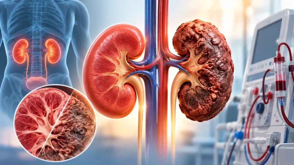
Kidney disorders can affect people of all ages and may develop silently over time. Many kidney problems do not cause clear symptoms in the early stages, which is why imaging tests play an important role in diagnosis and monitoring. One of the advanced imaging options available today is MRI, or Magnetic Resonance Imaging. MRI for kidney disorders uses magnetic fields and radio waves to create detailed pictures of the kidneys, surrounding tissues, and blood vessels without using harmful radiation.
MRI is often used when doctors need more detailed information than an ultrasound or X-ray can provide. It can help identify cysts, tumors, blockages, structural problems, blood vessel issues, and complications of kidney disease. In some cases, a special contrast dye is used to improve image clarity, but this must be used carefully in people with reduced kidney function.
What is MRI for Kidney Disorders?
MRI for kidney disorders is a non-invasive imaging test that uses strong magnetic fields and radio waves to create detailed images of the kidneys and nearby structures. Unlike X-ray or CT scan, MRI does not use ionizing radiation, which makes it a safer option for repeated imaging in many patients.
The test can show the size, shape, and position of the kidneys. It can also detect cysts, masses, stones, blockages, scarring, inflammation, and blood vessel problems. In some cases, a contrast agent called gadolinium is injected into a vein to highlight blood flow and improve image detail. The decision to use contrast depends on the patient’s kidney function and the clinical question being asked.
MRI is often recommended when ultrasound or CT scan does not give enough information, or when a more detailed view is needed for planning treatment. It is commonly used by nephrologists, urologists, radiologists, and surgeons to guide diagnosis and care.
Why MRI is Used for Kidney Disorders
MRI is used for many reasons in kidney care. It provides detailed images that help doctors understand the structure and function of the kidneys.
Common reasons for kidney MRI
- Evaluation of kidney cysts and masses.
- Detection of kidney tumors or suspicious growths.
- Assessment of kidney stones and blockages.
- Imaging of renal arteries for narrowing or blockage.
- Evaluation of congenital or structural kidney problems.
- Assessment of scarring or damage from chronic disease.
- Planning before kidney surgery or biopsy.
- Monitoring of known kidney conditions over time.
- Evaluation of complications such as infection or bleeding.
Why MRI may be preferred
- No ionizing radiation, which is safer for repeated scans.
- Excellent soft tissue detail for cysts and masses.
- Good visualization of blood vessels and blood flow.
- Useful when ultrasound is unclear or CT is not suitable.
- Helpful in planning surgery or interventional procedures.
Types of Kidney MRI
There are different types of MRI scans used for kidney disorders depending on the clinical need.
1. Non-contrast MRI
This type of MRI does not use any contrast dye. It is often used when kidney function is reduced or when contrast is not safe.
- Useful for basic structural imaging.
- Can detect cysts, masses, and some blockages.
- Safer for patients with poor kidney function.
- May provide less detail for blood vessels or small lesions.
2. Contrast-enhanced MRI
This type uses a gadolinium-based contrast agent injected into a vein. It improves visibility of blood vessels, tumors, and areas of inflammation.
- Provides more detailed images.
- Useful for evaluating tumors and vascular problems.
- Requires assessment of kidney function before use.
- Not suitable for some patients with severe kidney impairment.
3. MR angiography
This is a specialized MRI technique focused on blood vessels.
- Used to evaluate renal artery narrowing or blockage.
- Helpful in suspected renovascular hypertension.
- Can assess blood flow to the kidneys.
- May be done with or without contrast depending on the case.
4. Functional MRI techniques
Some advanced MRI methods assess kidney function and tissue characteristics.
- Diffusion-weighted imaging to assess tissue changes.
- Perfusion imaging to evaluate blood flow.
- Useful in research and selected clinical cases.
- May help in assessing chronic kidney disease progression.
Benefits of MRI for Kidney Disorders
MRI offers several advantages over other imaging methods for kidney evaluation.
Key benefits
- No ionizing radiation, which is safer for repeated scans.
- High-resolution images of soft tissues and structures.
- Excellent for detecting cysts, masses, and tumors.
- Detailed view of blood vessels and blood flow.
- Useful for surgical planning and biopsy guidance.
- Can detect problems that ultrasound or X-ray may miss.
- Non-invasive and painless in most cases.
- Helps monitor disease progression over time.
Why early imaging matters
Early detection of kidney problems can prevent complications and improve outcomes. MRI can identify issues before they become severe, allowing timely treatment and better preservation of kidney function.
Who May Need a Kidney MRI?
MRI for kidney disorders may be recommended for people with certain symptoms, findings, or risk factors.
Common indications
- Blood in urine without clear cause.
- Persistent flank or back pain.
- Suspected kidney cyst or mass on ultrasound.
- Uncontrolled high blood pressure with suspected renal artery issue.
- Abnormal kidney function tests.
- Recurrent urinary tract infections with structural concern.
- Family history of kidney disease or tumors.
- Follow-up of known kidney cysts or masses.
- Pre-surgical planning for kidney procedures.
Higher-risk groups
- People with chronic kidney disease.
- Patients with diabetes or hypertension.
- Those with a family history of kidney tumors.
- Individuals with recurrent stones or infections.
- Patients with abnormal imaging on ultrasound or CT.
- People with unexplained hematuria or proteinuria.
How is a Kidney MRI Performed?
The procedure is usually straightforward and takes about thirty to sixty minutes depending on the type of scan.
Before the scan
- Patient changes into a hospital gown.
- Metal objects such as jewelry, watches, and belts are removed.
- Medical history and implants are reviewed for safety.
- If contrast is planned, kidney function tests may be checked.
- Patient is positioned on a movable table that slides into the MRI machine.
During the scan
- Patient must lie still to get clear images.
- The machine makes loud tapping or knocking sounds.
- Earplugs or headphones are provided for comfort.
- If contrast is used, it is injected through a small IV line.
- The radiologist may ask the patient to hold breath briefly at times.
After the scan
- Patient can usually resume normal activities.
- If contrast was used, drinking water helps flush it out.
- Results are reviewed by a radiologist and shared with the treating doctor.
- Follow-up appointment may be scheduled to discuss findings.
Preparation for a Kidney MRI
Preparation depends on whether contrast is used and the patient’s overall health.
General preparation
- Inform the team about any implants, pacemakers, or metal devices.
- Share history of allergies, especially to contrast dye.
- Mention pregnancy or possibility of pregnancy.
- Disclose kidney disease or previous contrast reactions.
- Follow fasting instructions if given.
- Arrive early to complete paperwork and safety checks.
For contrast MRI
- Kidney function test may be required beforehand.
- Patient may be asked to avoid eating for a few hours.
- Hydration instructions may be given.
- Consent form may need to be signed.
- Allergy history must be shared clearly.
For patients with implants
- Most modern implants are MRI-safe, but confirmation is needed.
- Pacemakers and some metal devices may require special protocols.
- The radiology team will assess safety before the scan.
Safety and Risks of Kidney MRI
MRI is generally safe, but there are some considerations to keep in mind.
Safety points
- No ionizing radiation, which reduces long-term risk.
- Safe for most people, including those needing repeated imaging.
- Contrast use requires careful assessment in kidney disease.
- Gadolinium contrast is generally safe when kidney function is adequate.
- Rare allergic reactions to contrast can occur.
Possible risks
- Discomfort from lying still for a long time.
- Anxiety or claustrophobia in some patients.
- Loud noise during the scan.
- Rare allergic reaction to contrast dye.
- Risk of nephrogenic systemic fibrosis in severe kidney impairment with certain contrast agents.
- Movement can blur images and require repeat scanning.
Who may need extra caution
- Patients with severe kidney failure.
- Pregnant women, especially in the first trimester.
- People with certain implants or devices.
- Those with a history of contrast allergy.
- Patients with claustrophobia who may need sedation or open MRI.
Understanding MRI Results
MRI results are interpreted by a radiologist and shared with the treating doctor.
What results may show
- Normal kidney structure and size.
- Presence of simple cysts, which are usually benign.
- Complex cysts or masses that may need further evaluation.
- Kidney stones or blockages.
- Narrowing or blockage of renal arteries.
- Scarring or damage from chronic disease.
- Signs of infection, inflammation, or bleeding.
- Structural abnormalities present from birth.
What happens after results
- Doctor discusses findings with the patient.
- Further tests or biopsy may be recommended if needed.
- Treatment plan is made based on the diagnosis.
- Follow-up imaging may be scheduled to monitor the condition.
MRI vs Other Kidney Imaging Tests
Different imaging tests are used for different purposes in kidney care.
Ultrasound
- First-line, safe, and widely available.
- Good for basic screening and cyst detection.
- Less detailed for complex masses or vessels.
- No radiation or contrast needed.
CT scan
- Uses X-rays and may use contrast dye.
- Good for stones and acute problems.
- Higher radiation exposure than MRI.
- May not be ideal for repeated scans.
MRI
- No ionizing radiation.
- Excellent soft tissue and vascular detail.
- Useful for complex cases and surgical planning.
- Longer scan time and higher cost in some settings.
Why the choice matters
The right test depends on the clinical question. MRI is often chosen when detailed soft tissue or vascular imaging is needed, or when radiation exposure must be avoided.
Follow-Up Care After Kidney MRI
Follow-up depends on the findings and the underlying condition.
Follow-up pointers
- Review results with the treating doctor.
- Discuss any further tests or treatment needed.
- Monitor kidney function if contrast was used.
- Schedule repeat imaging if recommended.
- Follow lifestyle and medical advice for kidney health.
Why follow-up matters
Some kidney findings need monitoring over time. Regular follow-up ensures that any changes are detected early and managed promptly. It also helps assess the effectiveness of treatment.
Why Choose Rama Hospital for Kidney MRI?
Kidney disorders require careful evaluation and expert interpretation of imaging. A hospital with advanced MRI technology, experienced radiologists, and nephrology support can make a major difference in diagnosis and care.
- Advanced MRI machines for high-quality images.
- Experienced radiologists for accurate reporting.
- Safe use of contrast with proper kidney function assessment.
- Coordination between nephrology, urology, and radiology.
- Comprehensive care from diagnosis to treatment planning.
Frequently Asked Questions (FAQs)
Is kidney MRI safe?
Yes, MRI is generally safe and does not use ionizing radiation. However, contrast dye use requires careful assessment, especially in people with reduced kidney function. The radiology team will review safety before the scan.
Do I need contrast for a kidney MRI?
Not always. Contrast is used when more detailed imaging of blood vessels or tumors is needed. If kidney function is poor, the doctor may choose a non-contrast MRI to avoid risks.
How long does a kidney MRI take?
A kidney MRI usually takes thirty to sixty minutes. The exact time depends on whether contrast is used and how many images are needed. Patients need to lie still during the scan for clear images.
Can I eat before a kidney MRI?
For a non-contrast MRI, eating is usually allowed. For contrast MRI, fasting for a few hours may be required. The hospital will give clear instructions before the appointment.
Is MRI better than CT scan for kidneys?
MRI is better for soft tissue detail and does not use radiation, making it safer for repeated scans. CT is often better for detecting stones and acute problems. The choice depends on the clinical question and patient condition.
Can MRI detect kidney cancer?
Yes, MRI can detect kidney masses and tumors with good detail. It is often used to characterize lesions found on ultrasound or CT and to plan surgery if needed. Further testing may be required for confirmation.
What should I do after a contrast MRI?
Drink plenty of water to help flush the contrast dye from the body. Watch for any signs of allergic reaction such as rash, itching, or difficulty breathing, and seek help if these occur. Follow any specific instructions given by the hospital.
Conclusion
MRI for kidney disorders is a valuable imaging tool that provides detailed, radiation-free pictures of the kidneys and surrounding structures. It helps detect cysts, tumors, blockages, vascular problems, and structural issues that may not be clear on other tests. With proper preparation and safety checks, MRI is a safe and effective option for many patients.
The key to good outcomes is timely imaging, accurate interpretation, and appropriate follow-up. For patients with blood in urine, unexplained pain, abnormal kidney tests, or suspected masses, MRI can provide the clarity needed for diagnosis and treatment planning. With expert care and modern technology, kidney disorders can be detected early and managed effectively.
Rama Hospital can help evaluate kidney concerns, perform advanced MRI imaging when needed, and provide a clear treatment plan that supports kidney health and long-term well-being.