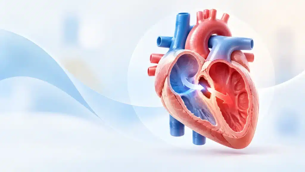
Ventricular Septal Defect, commonly called VSD, is one of the most common congenital heart defects seen in children and sometimes in adults. It is a hole in the wall, called the septum, that separates the two lower chambers of the heart, known as the ventricles. Because of this hole, oxygen-rich blood can flow from the left side of the heart into the right side, causing extra blood to go to the lungs. Over time, this can affect the heart, lungs, growth, and overall health of the child.
Many parents worry when they hear that their child has a heart defect. The good news is that Ventricular Septal Defect is often treatable, and many small defects close on their own as the child grows. Some children need medicines to support the heart and lungs, while others may need surgery or a catheter-based procedure to close the defect. The treatment depends on the size, location, and effect of the hole on the heart and lungs.
What is Ventricular Septal Defect?
A Ventricular Septal Defect is a hole in the wall between the two lower chambers of the heart. In a normal heart, the left ventricle pumps oxygen-rich blood to the body, and the right ventricle pumps oxygen-poor blood to the lungs. The wall between them, called the ventricular septum, keeps these two types of blood separate.
When there is a Ventricular Septal Defect, blood flows from the higher-pressure left ventricle into the lower-pressure right ventricle. This is called a left-to-right shunt. The extra blood then goes to the lungs, which can cause increased lung blood flow, lung congestion, and extra work for the heart.
The size of the hole matters a lot. A small Ventricular Septal Defect may cause no symptoms and may close on its own. A large VSD can cause breathing problems, poor feeding, slow growth, and heart failure if not treated. In adults, an untreated VSD can lead to lung damage and other complications.
Types of Ventricular Septal Defect
Ventricular Septal Defect can be classified based on where the hole is located in the ventricular septum.
Based on location
- Perimembranous VSD, which is the most common type and occurs near the valves at the top of the septum.
- Muscular VSD, which occurs in the lower, muscular part of the septum and is often small.
- Inlet VSD, which occurs near the valves that bring blood into the ventricles.
- Outlet or supracristal VSD, which occurs near the valves that send blood out of the heart.
Based on size
- Small VSD, which may cause no symptoms and often closes on its own.
- Moderate VSD, which may cause mild symptoms and needs monitoring.
- Large VSD, which can cause significant symptoms and may need treatment.
Based on direction of blood flow
- Left-to-right shunt, which is the most common pattern in VSD.
- Right-to-left shunt, which can happen in severe or complex cases and may cause low oxygen levels.
- Bidirectional shunt, where blood flows in both directions depending on pressure changes.
Causes of Ventricular Septal Defect
Ventricular Septal Defect is usually present from birth. It happens during early heart development when the wall between the ventricles does not close completely.
Common causes and contributing factors
- Genetic factors and family history of congenital heart defects.
- Chromosomal conditions such as Down syndrome.
- Maternal illness during pregnancy, such as uncontrolled diabetes.
- Certain infections during pregnancy.
- Exposure to harmful substances, alcohol, or certain medicines during pregnancy.
- Unknown causes in many cases.
Why the hole forms
During fetal development, the heart forms from simple structures into a complex four-chambered organ. If the septum does not close fully by the time of birth, a hole remains. This hole may be small or large, and its effect depends on size and location.
Risk Factors for VSD
Some factors increase the chance of a child being born with Ventricular Septal Defect.
Higher-risk factors
- Family history of congenital heart disease.
- Maternal diabetes, especially if poorly controlled.
- Maternal infections during early pregnancy.
- Use of certain medicines or alcohol during pregnancy.
- Exposure to harmful chemicals or radiation.
- Chromosomal abnormalities in the baby.
- Advanced maternal age in some cases.
Why risk increases
These factors can interfere with normal heart development in the fetus. While many cases happen without any known cause, awareness of risk factors helps in early detection and planning.
Symptoms of VSD
Symptoms depend on the size of the hole and the amount of extra blood going to the lungs.
Symptoms in infants
- Fast breathing or breathlessness.
- Poor feeding or tiredness during feeding.
- Slow weight gain or poor growth.
- Frequent respiratory infections.
- Sweating during feeds.
- Irritability.
- Cyanosis or bluish tint in severe cases.
Symptoms in older children
- Fatigue.
- Shortness of breath during activity.
- Frequent chest infections.
- Poor exercise tolerance.
- Heart murmur detected during examination.
- Slow growth compared to peers.
Symptoms in adults
- Breathlessness.
- Fatigue.
- Palpitations.
- Reduced exercise capacity.
- Signs of heart failure in untreated large defects.
- Arrhythmias or irregular heartbeat in some cases.
Heart murmur
Many children with VSD have a heart murmur, which is an extra sound heard during a heartbeat. This murmur is often the first clue that leads to further testing. Not all murmurs mean a serious problem, but they should be evaluated by a cardiologist.
VSD in Different Age Groups
Ventricular Septal Defect can present differently depending on age.
In newborns and infants
- Often detected through murmur or symptoms like poor feeding and fast breathing.
- Small defects may be silent and found later.
- Large defects can cause early heart failure signs.
In children
- May be found during routine check-up due to murmur.
- Some children have no symptoms and grow normally.
- Larger defects may affect growth and activity level.
In adults
- May be an untreated childhood defect.
- Can cause complications such as arrhythmia, valve problems, or lung damage.
- Some adults are diagnosed incidentally during tests for other issues.
How is VSD Diagnosed?
Diagnosis is based on symptoms, physical examination, and imaging tests. A cardiologist uses these tools to assess the size, location, and effect of the defect.
Diagnostic steps
- Detailed history of symptoms and growth.
- Family history of heart defects.
- Physical examination, especially listening for a murmur.
- Assessment of oxygen levels and heart function.
- Imaging tests to visualize the heart structure.
Common tests that may be done
- Echocardiogram, which is the main test to see the hole and measure blood flow.
- Electrocardiogram to check heart rhythm.
- Chest X-ray to assess heart size and lung congestion.
- Pulse oximetry to measure oxygen levels.
- Cardiac MRI in selected cases for detailed anatomy.
- Cardiac catheterization in complex cases or before intervention.
Why diagnosis matters
Early and accurate diagnosis helps decide whether the Ventricular Septal Defect needs monitoring, medicines, or closure. It also helps prevent complications such as heart failure, lung damage, or infection of the heart.
Treatment for VSD
Treatment depends on the size, location, symptoms, and effect on the heart and lungs.
1. Observation and monitoring
Many small VSDs close on their own, especially in infants.
- Regular follow-up with a cardiologist.
- Echocardiogram at intervals to check size and closure.
- Monitoring growth and symptoms.
- No treatment needed if the defect is small and not causing problems.
2. Medicines
Medicines may be used to support the heart and lungs while waiting for closure or before surgery.
- Diuretics to reduce fluid buildup.
- Medicines to improve heart function.
- Medicines to control symptoms of heart failure.
- Antibiotics before certain procedures to prevent infection of the heart.
3. Nutritional support
For infants with poor feeding or slow growth, nutrition support may be needed.
- High-calorie feeds.
- Smaller, more frequent feeds.
- Feeding tubes in severe cases.
- Close monitoring of weight and growth.
4. Catheter-based closure
Some VSDs can be closed using a catheter inserted through a blood vessel, without open-heart surgery.
- A device is placed to seal the hole.
- Minimally invasive with shorter recovery.
- Suitable for certain types and sizes of Ventricular Septal Defect.
- May not be possible for all defects.
5. Surgical closure
Open-heart surgery may be needed for large VSDs or those not suitable for catheter closure.
- The surgeon closes the hole with a patch or stitches.
- Performed under general anesthesia.
- Requires a hospital stay and recovery period.
- Highly effective with good long-term outcomes.
6. Treatment of complications
If complications such as heart failure, arrhythmia, or valve problems occur, additional treatment may be needed.
- Medicines to manage heart failure.
- Procedures for valve repair if needed.
- Treatment for irregular heartbeat.
- Long-term follow-up to monitor heart and lung health.
Recovery After Treatment
Recovery depends on the type of treatment and the size of the defect.
Recovery after observation
- Regular follow-up appointments.
- Monitoring for closure or change in size.
- Normal growth and activity if the defect is small.
Recovery after catheter closure
- Shorter hospital stay.
- Quick return to normal activities in many cases.
- Follow-up imaging to confirm closure.
- Medicines may be needed temporarily.
Recovery after surgery
- Longer hospital stay, often in intensive care initially.
- Gradual return to feeding and activity.
- Wound care and monitoring.
- Rehabilitation and follow-up to ensure heart function is normal.
- Most children recover well and lead normal lives.
Possible Complications
If Ventricular Septal Defect is not treated or is large, complications can occur.
Possible complications
- Heart failure due to extra workload.
- Pulmonary hypertension or high lung pressure.
- Irregular heartbeat.
- Valve problems.
- Infection of the heart lining.
- Slow growth and poor feeding in infants.
- Lung damage over time.
- Reduced exercise capacity.
- Cyanosis in severe cases.
Why complications happen
Extra blood flow to the lungs and heart can cause strain and damage over time. Early detection and treatment help prevent these complications.
When to See a Doctor?
Medical evaluation is important if symptoms suggest a heart problem.
- Fast breathing or breathlessness.
- Poor feeding or sweating during feeds.
- Slow weight gain.
- Frequent chest infections.
- Bluish tint to lips or skin.
- Fatigue or reduced activity.
- Heart murmur detected during check-up.
Prevention of Complications
While Ventricular Septal Defect itself may not be preventable, complications can be reduced.
Prevention pointers
- Early diagnosis and regular follow-up.
- Timely treatment of symptoms.
- Good nutrition and growth monitoring.
- Prevention of infections with good hygiene.
- Antibiotic prophylaxis before certain procedures if advised.
- Control of other risk factors such as blood pressure in adults.
- Avoid smoking and alcohol exposure during pregnancy.
For parents
- Attend all follow-up appointments.
- Watch for warning signs.
- Follow feeding and medication advice.
- Ensure vaccinations are up to date.
Why Choose Rama Hospital for VSD Treatment?
Ventricular Septal Defect requires expert pediatric cardiology, advanced imaging, and sometimes surgical care. A hospital with experienced cardiologists, pediatric surgeons, and intensive care support can make a major difference in outcomes.
- Expert evaluation by pediatric cardiologists and cardiac surgeons.
- Advanced echocardiography and imaging.
- Catheter-based and surgical closure options.
- Intensive care support for complex cases.
- Long-term follow-up and rehabilitation.
- Family education and support.
Frequently Asked Questions (FAQs)
Is Ventricular Septal Defect serious?
It depends on the size and location. Small VSDs may cause no problems and close naturally. Large VSDs can cause heart failure and lung damage if not treated.
Can VSD close on its own?
Yes, many small VSDs close on their own during infancy or early childhood. Regular follow-up with a cardiologist is needed to monitor this.
How is VSD treated?
Treatment may include observation, medicines, catheter-based closure, or surgery. The choice depends on size, symptoms, and effect on the heart.
Can adults have VSD?
Yes, some adults have untreated Ventricular Septal Defect from childhood. It can cause complications such as arrhythmia or lung problems, so evaluation is important.
What are the signs of VSD in babies?
Signs include fast breathing, poor feeding, sweating during feeds, slow weight gain, and frequent infections. A heart murmur may also be heard.
Is surgery always needed for VSD?
No, surgery is not always needed. Small VSDs may only need monitoring. Larger or symptomatic defects may need catheter closure or surgery.
Can a child with VSD live a normal life?
Yes, many children with Ventricular Septal Defect live normal, active lives, especially after closure or if the defect is small. Regular follow-up helps ensure long-term heart health.
Conclusion
Ventricular Septal Defect is a common congenital heart defect that affects the wall between the heart’s lower chambers. While many small VSDs close on their own, larger defects can cause serious problems if not treated. Early diagnosis, regular monitoring, and timely treatment are key to preventing complications.
The most important steps are proper evaluation with echocardiography, follow-up with a cardiologist, and treatment when needed. With modern cardiology and surgical care, most children and adults with VSD can lead healthy, active lives. Rama Hospital can provide expert evaluation, advanced treatment options, and long-term support for patients with Ventricular Septal Defect.