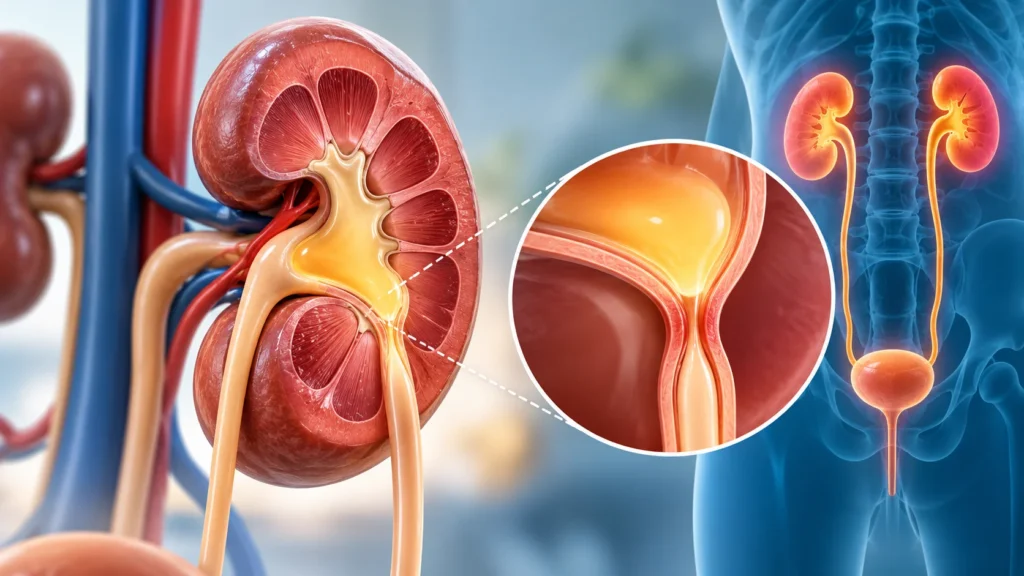
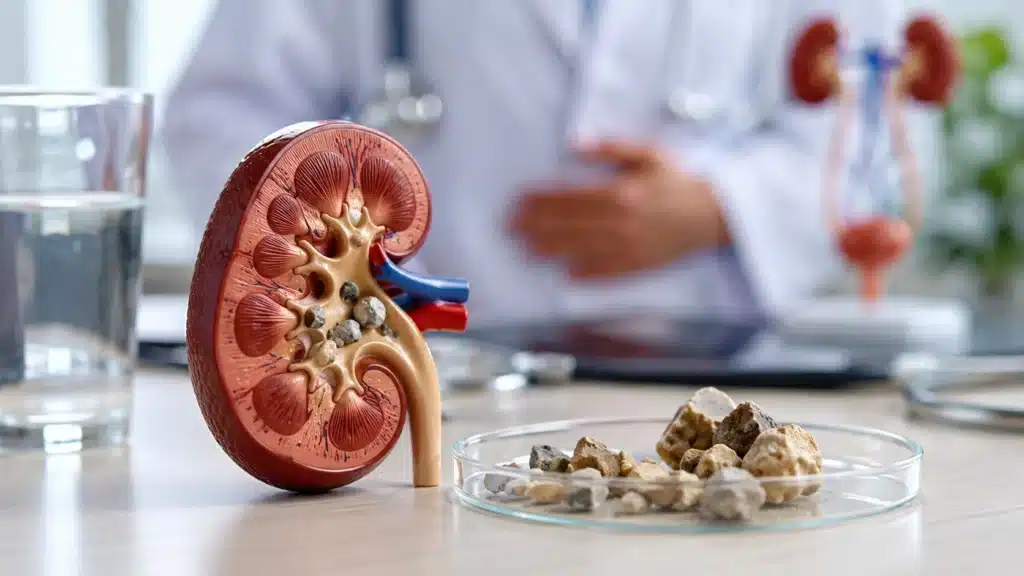
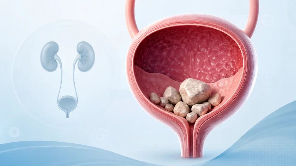
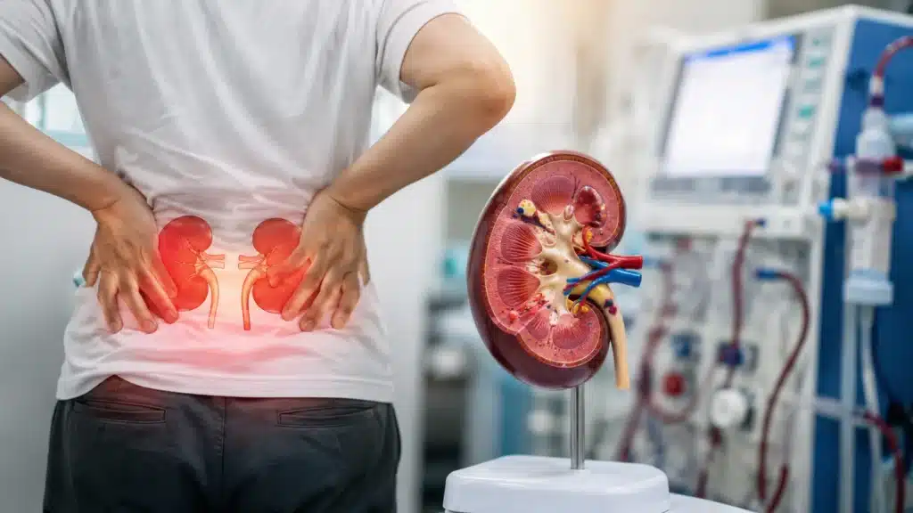
Ureteropelvic junction obstruction, commonly called UPJ obstruction, is a condition where the flow of urine from the kidney to the ureter is blocked or narrowed. The ureteropelvic junction is the point where the renal pelvis of the kidney meets the ureter, which carries urine to the bladder. When this area is narrowed, urine cannot drain properly. This causes urine to back up into the kidney, leading to swelling of the kidney, known as hydronephrosis.
UPJ obstruction can be present from birth or develop later in life. In many cases, it is detected during pregnancy or in early childhood. In adults, it may be discovered during evaluation for recurrent urinary infections, kidney stones, flank pain, or abnormal imaging. If left untreated, long-standing obstruction can damage the kidney, reduce its function, and increase the risk of infections and stones.
What is Ureteropelvic Junction Obstruction?
UPJ obstruction is a blockage or narrowing at the junction between the kidney and the ureter. This prevents urine from flowing smoothly from the renal pelvis into the ureter. As a result, urine builds up in the kidney, causing it to swell. This swelling is called hydronephrosis.
The obstruction can be partial or complete. In partial obstruction, some urine can still pass, but not enough to prevent swelling. In complete obstruction, urine flow is severely restricted. Over time, the pressure from the backed-up urine can stretch the kidney tissue and affect its function.
UPJ obstruction is one of the most common causes of hydronephrosis detected before birth. It can affect one kidney or both kidneys. Many children with mild obstruction improve on their own. Others may need surgery to correct the narrowing and protect kidney function.
Types of UPJ Obstruction
UPJ obstruction can be classified based on when it occurs, whether it affects one or both kidneys, and the underlying cause.
Based on timing
- Congenital UPJ obstruction, which is present from birth due to abnormal development of the ureteropelvic junction.
- Acquired UPJ obstruction, which develops later in life due to scarring, stones, surgery, or other conditions.
Based on involvement
- Unilateral UPJ obstruction, which affects one kidney.
- Bilateral UPJ obstruction, which affects both kidneys and may carry a higher risk of kidney function problems.
Based on cause
- Intrinsic obstruction, where the narrowing is within the wall of the ureter or junction itself.
- Extrinsic obstruction, where something outside the ureter, such as a crossing blood vessel, presses on the junction and causes narrowing.
Causes of UPJ Obstruction
The cause of UPJ obstruction depends on whether it is congenital or acquired.
Congenital causes
- Abnormal development of the ureteropelvic junction during fetal growth.
- Lack of proper muscle fibers in the wall of the ureter at the junction.
- Narrowing of the ureter at the junction without a clear external cause.
- Abnormal positioning of blood vessels that cross and compress the ureter.
Acquired causes
- Scarring from previous kidney or ureter surgery.
- Scarring after kidney stones or repeated infections.
- Inflammation or fibrosis around the ureter.
- Trauma to the kidney or ureter area.
- Rare tumors or growths pressing on the junction.
Why obstruction happens
In congenital cases, the ureter may not develop the right structure to allow smooth urine flow. In acquired cases, injury, inflammation, or scarring can narrow the passage. In some patients, a blood vessel crosses over the ureter and compresses it, causing functional obstruction.
Risk Factors for UPJ Obstruction
Some people are more likely to develop ureteropelvic junction obstruction than others.
Higher-risk groups
- Fetuses detected with hydronephrosis on prenatal ultrasound.
- Newborns and infants with urinary tract abnormalities.
- Children with a history of recurrent urinary infections.
- Patients with a history of kidney stones.
- Individuals who have had previous kidney or ureter surgery.
- People with structural abnormalities of the urinary tract.
Why risk increases in these groups
- Developmental issues during pregnancy can affect ureter formation.
- Recurrent infections or stones can cause scarring.
- Previous surgery can lead to narrowing at the junction.
- Anatomical variations may predispose certain individuals to obstruction.
Symptoms of UPJ Obstruction
Symptoms vary depending on age, severity of obstruction, and whether one or both kidneys are affected. Many people, especially children, may have no symptoms at all.
Symptoms in infants and children
- Detected as hydronephrosis on prenatal or postnatal ultrasound.
- Recurrent urinary tract infections.
- Fever without clear source.
- Poor feeding or failure to thrive in severe cases.
- Abdominal or flank pain in older children.
- Vomiting or irritability in infants.
- Palpable abdominal mass in some cases.
Symptoms in adults
- Flank pain on one side, often intermittent.
- Pain that worsens after drinking large amounts of fluid.
- Recurrent urinary tract infections.
- Blood in urine.
- Kidney stones.
- Nausea or vomiting during pain episodes.
- High blood pressure in some cases due to kidney involvement.
- Reduced kidney function detected on blood tests.
Emergency warning signs
- Severe flank pain with fever.
- High fever with chills and flank pain.
- Inability to pass urine.
- Severe vomiting with pain.
- Signs of infection such as confusion, rapid heartbeat, or low blood pressure.
UPJ Obstruction in Different Age Groups
The presentation and management of ureteropelvic junction obstruction can differ between children and adults.
In infants and children
- Often detected during routine prenatal ultrasound.
- Many mild cases resolve on their own without surgery.
- Severe cases may need early surgical correction to protect kidney function.
- Recurrent infections are a common reason for evaluation.
In adults
- May be discovered during evaluation for flank pain, stones, or infections.
- Symptoms may have been present for years but overlooked.
- Surgery is often recommended if symptoms are significant or kidney function is at risk.
- Adults may have additional factors such as stones or scarring.
How is UPJ Obstruction Diagnosed?
Diagnosis is based on symptoms, physical examination, and imaging tests. Since many patients have no symptoms, UPJ obstruction is often found incidentally during tests for other reasons.
Diagnostic steps
- Detailed history of symptoms such as pain, infections, or stones.
- Family history of urinary tract abnormalities.
- Physical examination, including abdominal and flank assessment.
- Urine tests to check for infection or blood.
- Blood tests to assess kidney function.
- Imaging tests to visualize the kidneys and ureters.
Common tests that may be done
- Ultrasound of the kidneys and bladder to detect hydronephrosis.
- CT scan or MR urography to see the urinary tract in detail.
- Diuretic renal scan to assess kidney function and drainage.
- Voiding cystourethrogram in children to rule out other urinary abnormalities.
- Intravenous pyelogram in selected cases.
- Blood tests such as creatinine and urea to evaluate kidney function.
Why diagnosis matters
Early diagnosis helps prevent kidney damage. Imaging can show the degree of swelling, the location of obstruction, and the function of each kidney. This information guides whether observation or surgery is the best option.
Treatment for UPJ Obstruction
Treatment depends on the age of the patient, severity of obstruction, symptoms, kidney function, and whether complications such as infections or stones are present.
Treatment for mild cases
- Observation with regular ultrasound monitoring.
- Antibiotics to prevent or treat urinary infections.
- Monitoring of kidney function with blood tests.
- Many infants with mild hydronephrosis improve without surgery.
Treatment for moderate to severe cases
- Surgical correction is often recommended if obstruction is significant.
- Surgery aims to remove the narrowed segment and reconstruct the junction.
- The goal is to improve urine flow and protect kidney function.
- Surgery may be recommended earlier if there are recurrent infections, pain, stones, or declining kidney function.
1. Pyeloplasty
Pyeloplasty is the standard surgical treatment for ureteropelvic junction obstruction. It involves removing the narrowed segment of the ureter and reconnecting the healthy ureter to the renal pelvis.
- Can be done as open surgery, laparoscopic surgery, or robotic-assisted surgery.
- Minimally invasive approaches are common in adults and older children.
- Success rates are high, with most patients experiencing improved drainage and symptom relief.
- A temporary stent may be placed to support healing.
2. Endopyelotomy
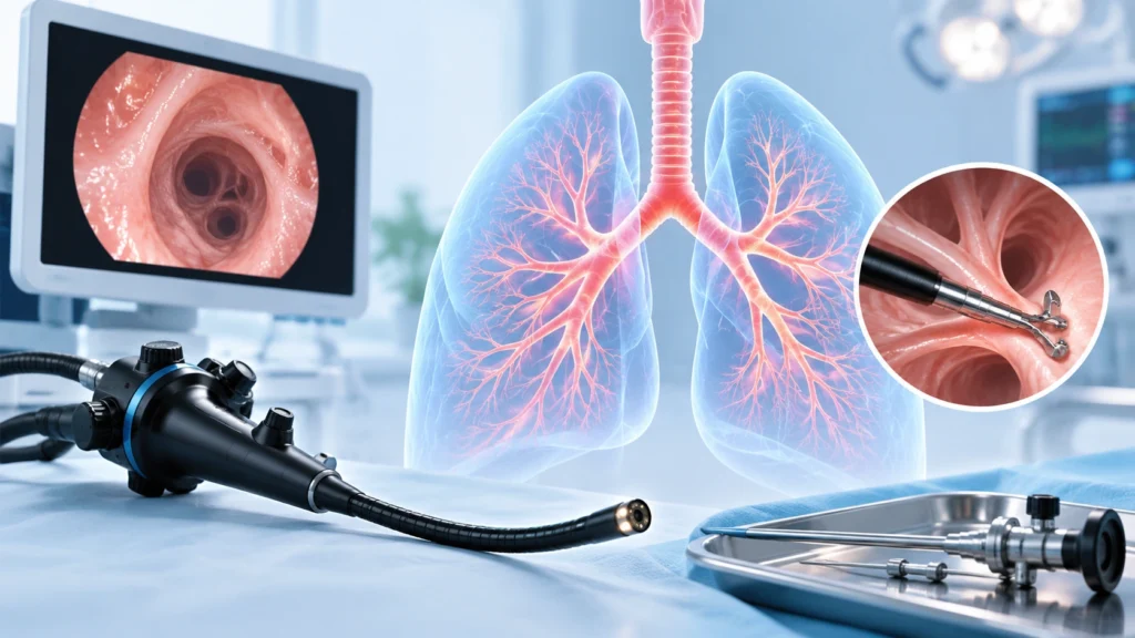
Endopyelotomy is a less invasive option where the narrowed area is cut from inside using a scope.
- Usually done through the urethra or through a small puncture in the back.
- May be suitable for selected adult patients.
- Success rates are lower than pyeloplasty in some cases.
- May not be ideal for children or complex obstructions.
3. Stent placement
In some cases, a ureteral stent may be placed temporarily to improve drainage.
- Used as a bridge to surgery or in patients who are not immediate surgical candidates.
- Helps relieve obstruction and protect kidney function.
- Requires follow-up for removal or replacement.
4. Nephrostomy tube
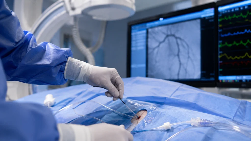
In severe cases with infection or poor drainage, a tube may be placed directly into the kidney through the back.
- Drains urine externally to relieve pressure.
- Used in emergency situations or before definitive surgery.
- Temporary measure until the patient is stable for reconstruction.
5. Supportive care
For patients with infections or stones, additional treatment is needed.
- Antibiotics for urinary infections.
- Stone removal procedures if stones are present.
- Pain management during acute episodes.
- Monitoring of kidney function over time.
Recovery After Treatment
Recovery depends on the type of treatment, age of the patient, and whether complications were present.
Recovery after pyeloplasty
- Hospital stay is usually short for minimally invasive surgery.
- Pain is managed with medicines.
- A stent may be placed and removed later in a simple procedure.
- Most patients resume normal activities within a few weeks.
- Follow-up imaging is done to confirm improved drainage.
Recovery after endoscopic procedures
- Shorter recovery time compared to open surgery.
- May require stent placement temporarily.
- Follow-up imaging to ensure success.
- Some patients may need additional treatment if obstruction recurs.
Long-term follow-up
- Regular ultrasound or renal scan to monitor kidney drainage.
- Blood tests to check kidney function.
- Monitoring for recurrent infections or stones.
- Continued care with a urologist as needed.
Possible Complications
Ureteropelvic junction obstruction, especially if untreated, can lead to several complications.
Complications of untreated obstruction
- Progressive kidney damage.
- Loss of kidney function.
- Recurrent urinary tract infections.
- Kidney stone formation.
- High blood pressure due to kidney involvement.
- Chronic flank pain.
- Scarring of kidney tissue.
Complications after treatment
- Infection at the surgical site.
- Bleeding.
- Stent-related discomfort or infection.
- Recurrence of obstruction in rare cases.
- Need for additional procedures if drainage is not adequate.
Why complications happen
Long-standing obstruction increases pressure inside the kidney, which can damage delicate kidney tissue. Infections and stones can further worsen kidney health. Timely treatment reduces the risk of permanent damage.
When to See a Doctor
Medical evaluation is important for people with symptoms or risk factors suggestive of UPJ obstruction.
- Recurrent flank pain on one side.
- Recurrent urinary tract infections.
- Blood in urine.
- History of kidney stones.
- Hydronephrosis detected on ultrasound.
- Poor kidney function on blood tests.
- Abdominal mass or swelling in a child.
Prevention of UPJ Obstruction
Congenital ureteropelvic junction obstruction cannot be prevented, but complications can be reduced with early detection and treatment.
Prevention pointers
- Regular prenatal ultrasound during pregnancy.
- Postnatal follow-up for infants with hydronephrosis.
- Prompt treatment of urinary infections.
- Adequate fluid intake to reduce stone risk.
- Regular check-ups for patients with known urinary abnormalities.
- Early urology evaluation for recurrent symptoms.
For children with hydronephrosis
- Follow pediatric urology advice for monitoring.
- Complete all recommended imaging and tests.
- Watch for signs of infection or pain.
- Maintain regular follow-up even if symptoms are mild.
Why Choose Rama Hospital for UPJ Obstruction Treatment?
Ureteropelvic junction obstruction requires expert urological evaluation, advanced imaging, and often reconstructive surgery. A hospital with experienced urologists, pediatric urology support, and modern surgical technology can provide comprehensive care.
- Expert evaluation by urology and pediatric urology specialists.
- Advanced imaging for accurate diagnosis.
- Minimally invasive and open surgical options.
- Care for both children and adults.
- Management of complications such as infections and stones.
- Long-term follow-up to protect kidney function.
Frequently Asked Questions (FAQs)
Is ureteropelvic junction obstruction serious?
It can be serious if it causes significant blockage, recurrent infections, or kidney damage. Many mild cases in children improve on their own. Severe cases need treatment to protect kidney function and prevent complications.
Can UPJ obstruction be cured?
Yes, most cases can be treated effectively. Surgery such as pyeloplasty has high success rates in relieving obstruction and improving kidney drainage. Early treatment helps prevent long-term kidney damage.
What are the symptoms of ureteropelvic junction obstruction?
Symptoms may include flank pain, recurrent urinary infections, blood in urine, kidney stones, or hydronephrosis detected on ultrasound. Many children have no symptoms and are diagnosed through imaging.
How is ureteropelvic junction obstruction diagnosed?
Diagnosis is done using ultrasound, CT or MR urography, and renal scan to assess kidney function and drainage. Urine and blood tests help check for infection and kidney function.
Does ureteropelvic junction obstruction always need surgery?
No, mild cases, especially in infants, may be monitored without surgery. Surgery is recommended if obstruction is severe, symptoms are present, kidney function is declining, or complications occur.
What is pyeloplasty?
Pyeloplasty is a surgical procedure to remove the narrowed segment of the ureter and reconstruct the junction between the kidney and ureter. It is the standard treatment for significant UPJ obstruction.
Can ureteropelvic junction obstruction come back after surgery?
Recurrence is uncommon but possible. Regular follow-up imaging helps ensure the kidney is draining well. If symptoms return, further evaluation may be needed.
Conclusion
Ureteropelvic junction obstruction is a condition that affects the flow of urine from the kidney to the ureter. It can lead to kidney swelling, infections, stones, and long-term kidney damage if not managed properly. The good news is that most cases can be diagnosed early and treated effectively with modern urological care.
The most important steps are proper diagnosis, timely treatment when needed, and long-term follow-up to protect kidney function. For patients with flank pain, recurrent infections, hydronephrosis, or abnormal kidney tests, a urology evaluation can make a major difference. With the right care, patients can preserve kidney health and prevent complications.
Rama Hospital can help evaluate UPJ obstruction, provide expert surgical and medical management, and offer comprehensive follow-up to ensure the best possible outcomes for children and adults.