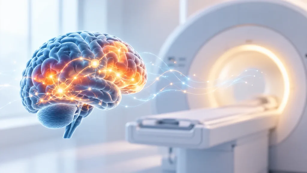
Functional MRI, commonly known as fMRI, is an advanced imaging technique that helps doctors see how the brain is working in real time. Unlike a standard MRI, which shows the structure of the brain, fMRI shows brain activity by measuring changes in blood flow. When a part of the brain is active, it uses more oxygen, and blood flow to that area increases. fMRI can detect these changes and create detailed maps of brain function.
This technology is especially useful in neurology, neurosurgery, psychiatry, and rehabilitation. It helps doctors plan surgery for brain tumors, evaluate epilepsy, study stroke recovery, understand memory and language areas, and assess certain neurological and psychiatric conditions. For patients with complex brain disorders, fMRI provides information that cannot be obtained from routine scans.
What is Functional MRI (fMRI)?
Functional MRI is a specialized type of magnetic resonance imaging that measures brain activity by detecting changes in blood flow. It is based on the principle that active brain areas receive more oxygenated blood. By tracking these changes, fMRI can show which parts of the brain are involved in specific tasks such as speaking, moving, thinking, or remembering.
fMRI does not use radiation. It uses strong magnetic fields and radio waves, similar to a standard MRI scanner. The difference is in how the images are processed. While a regular MRI shows anatomy, fMRI shows function. This makes it a powerful tool for understanding how the brain works in health and disease.
fMRI is often used before brain surgery to identify important areas such as language, motor, or memory regions. It is also used in research and clinical evaluation of conditions like epilepsy, stroke, dementia, traumatic brain injury, and certain psychiatric disorders.
How Does Functional MRI Work?
fMRI works by detecting changes in blood oxygen levels in different parts of the brain. When neurons in a specific brain area become active, they require more oxygen. The body responds by increasing blood flow to that region. This change in blood oxygenation can be measured by the MRI scanner.
Key principles of Functional MRI
- Active brain areas use more oxygen.
- Increased neural activity leads to increased blood flow.
- Oxygenated and deoxygenated blood have different magnetic properties.
- The MRI scanner detects these differences.
- Computer software creates maps showing which areas are active during specific tasks.
Common tasks during Functional MRI
- Moving fingers, toes, or limbs to map motor areas.
- Speaking or listening to map language areas.
- Looking at images or patterns to map visual areas.
- Performing memory or thinking tasks to map cognitive regions.
- Resting-state fMRI, where the patient lies still while the scanner measures baseline brain activity.
The result is a set of images that show both the structure of the brain and the areas that are active during different functions.
Common Uses of Functional MRI
fMRI has many clinical and diagnostic uses. It is particularly helpful when doctors need to understand how a disease or injury is affecting brain function.
Pre-surgical brain mapping
- Identifying language areas before tumor surgery.
- Mapping motor areas to avoid damage during surgery.
- Planning safe surgical approaches for epilepsy or lesions.
- Reducing the risk of post-surgical deficits.
Epilepsy evaluation
- Locating the seizure focus in the brain.
- Understanding how seizures affect brain networks.
- Planning surgical or device-based treatment for drug-resistant epilepsy.
Stroke assessment
- Evaluating brain recovery after stroke.
- Identifying which areas are still functional.
- Guiding rehabilitation strategies.
- Studying brain reorganization after injury.
Brain tumor evaluation
- Determining how close a tumor is to critical brain areas.
- Assessing whether important functions are displaced or affected.
- Helping neurosurgeons plan safer operations.
Neurological and psychiatric conditions
- Studying memory and cognition in dementia.
- Evaluating brain changes in traumatic brain injury.
- Research applications in depression, anxiety, and schizophrenia.
- Understanding brain networks in developmental disorders.
Rehabilitation and therapy planning
- Tracking recovery after brain injury.
- Guiding speech, motor, or cognitive therapy.
- Monitoring response to treatment over time.
Benefits of Functional MRI
fMRI offers several advantages over other imaging methods when it comes to brain function.
Non-invasive and safe
- No needles or injections are usually required.
- No ionizing radiation is used.
- Suitable for repeated studies when needed.
Detailed functional information
- Shows which brain areas are active during specific tasks.
- Helps distinguish between healthy and affected tissue.
- Provides information that structural scans cannot show.
Surgical planning support
- Reduces risk of damaging important brain areas.
- Helps surgeons choose the safest approach.
- Improves confidence in complex brain procedures.
Research and clinical insight
- Helps doctors understand how diseases affect brain networks.
- Supports development of new treatments.
- Provides objective data for complex neurological cases.
Who May Need Functional MRI?
Not every patient with a brain condition needs fMRI. It is usually recommended when detailed functional information will change management or improve safety.
Patients who may benefit
- People with brain tumors near language or motor areas.
- Patients being evaluated for epilepsy surgery.
- Stroke survivors with ongoing deficits.
- Individuals with traumatic brain injury and cognitive changes.
- Patients with complex neurological symptoms unexplained by routine scans.
- People participating in specialized rehabilitation programs.
When fMRI may not be needed
- Routine headache evaluation without neurological signs.
- Simple structural problems clearly seen on standard MRI.
- Cases where the result will not change treatment.
- Patients who cannot lie still or follow instructions during the scan.
The Functional MRI Procedure
The fMRI procedure is similar to a standard MRI but includes specific tasks or instructions to activate different brain areas.
Before the scan
- The patient is screened for metal implants, pacemakers, or other contraindications.
- Instructions are given about clothing, jewelry, and personal items.
- The purpose of the scan and the tasks are explained.
- The patient may practice simple tasks before entering the scanner.
During the scan
- The patient lies on a movable table that slides into the MRI machine.
- A head coil is placed around the head to improve image quality.
- The patient is asked to perform tasks such as moving fingers, speaking, or looking at images.
- In resting-state fMRI, the patient lies still with eyes open or closed.
- The scan is painless but can be noisy. Ear protection is provided.
- The patient must try to stay as still as possible for clear images.
After the scan
- The patient can usually resume normal activities immediately.
- A radiologist or specialist analyzes the images.
- Results are shared with the treating doctor.
- Further discussion may be needed to plan treatment or surgery.
Preparation for Functional MRI
Proper preparation helps ensure the scan is safe and produces good quality images.
Important preparation steps
- Inform the doctor about any metal implants, devices, or previous surgeries.
- Remove all metal objects, jewelry, and electronic devices before the scan.
- Wear comfortable clothing without metal zippers or buttons.
- Follow any specific instructions about food, drink, or medicines.
- Practice staying still for the duration of the scan.
- Inform the team if you feel anxious or claustrophobic.
Special considerations
- Children may need sedation or child-friendly preparation.
- Patients with severe anxiety may need support or mild sedation.
- People with hearing difficulties may need adjusted communication methods.
- Patients with movement disorders may require additional stabilization.
Safety and Limitations
fMRI is generally safe, but there are some important considerations.
Safety points
- No ionizing radiation is used.
- The magnetic field is strong, so metal objects must be kept away.
- People with certain implants or devices may not be eligible.
- The scanner is noisy, so ear protection is always provided.
- Some people may feel claustrophobic inside the machine.
Limitations of fMRI
- Requires the patient to lie still and follow instructions.
- Movement can reduce image quality.
- Not all brain functions can be mapped equally well.
- Results must be interpreted by experienced specialists.
- fMRI shows correlation, not direct cause, of brain activity.
Functional MRI in Neurosurgery and Epilepsy Care
One of the most important uses of fMRI is in planning brain surgery and managing epilepsy.
In neurosurgery
- Helps identify critical areas before removing tumors.
- Reduces risk of language or motor deficits after surgery.
- Guides the surgical approach to spare important tissue.
- Supports decision-making in complex cases.
In epilepsy
- Helps locate the seizure focus when other tests are unclear.
- Assists in planning resective surgery or device implantation.
- Evaluates how seizures affect brain networks.
- Supports long-term management strategies.
Functional MRI in Stroke and Rehabilitation
fMRI is increasingly used in stroke care and recovery programs.
In stroke assessment
- Shows which brain areas are still active after stroke.
- Helps predict recovery potential.
- Identifies compensatory brain activity.
- Guides targeted rehabilitation.
In rehabilitation
- Tracks changes in brain activity during therapy.
- Supports personalized recovery plans.
- Helps measure progress over time.
- Provides motivation through objective feedback.
Why Choose Rama Hospital for Functional MRI and Neuroimaging?
Functional MRI is a specialized test that requires advanced equipment, experienced radiologists, and close coordination with neurologists and neurosurgeons. A hospital with strong neuroimaging capabilities can provide more accurate diagnosis and better treatment planning.
- Access to advanced MRI and fMRI technology.
- Experienced radiologists and neuro specialists.
- Integrated care for brain tumors, epilepsy, stroke, and neurological disorders.
- Safe and patient-friendly imaging environment.
- Clear communication of results to patients and families.
Frequently Asked Questions (FAQs)
Is Functional MRI painful or dangerous?
No, fMRI is not painful and does not use radiation. It uses magnetic fields and radio waves like a standard MRI. The main challenges are staying still and tolerating the noise and confined space inside the scanner.
How long does an Functional MRI scan take?
An fMRI scan usually takes between thirty to sixty minutes, depending on the type of study and the tasks involved. The exact time will be explained by the imaging team before the scan.
Do I need any special preparation for Functional MRI?
You may need to avoid metal objects, wear comfortable clothing, and follow instructions about food or medicines. It is important to inform the team about any implants, devices, or anxiety issues before the scan.
Can children undergo Functional MRI?
Yes, children can have fMRI when needed. Younger children may require sedation or child-friendly preparation to help them stay still during the scan. The decision is made based on age and clinical need.
What conditions can Functional MRI help diagnose?
fMRI is commonly used for brain tumor planning, epilepsy evaluation, stroke assessment, traumatic brain injury, and certain neurological or psychiatric conditions. It is most useful when functional information will guide treatment.
Will Functional MRI change my treatment plan?
In many cases, yes. functional MRI can help surgeons plan safer operations, guide epilepsy treatment, support stroke rehabilitation, and clarify complex neurological symptoms. Your doctor will explain how the results may affect your care.
Is Functional MRI available at all hospitals?
No, functional MRI requires specialized MRI scanners and trained staff. It is available only at centers with advanced neuroimaging capabilities. Your doctor can refer you to an appropriate facility if fMRI is recommended.
Conclusion
Functional MRI is a powerful tool that helps doctors see how the brain works, not just how it looks. It provides critical information for brain surgery, epilepsy care, stroke recovery, and complex neurological conditions. While it is not needed for every patient, fMRI can make a major difference when precise brain mapping is required.
The test is safe, non-invasive, and does not use radiation. However, it requires cooperation, stillness, and expert interpretation. For patients with brain tumors, epilepsy, stroke, or unexplained neurological symptoms, fMRI can provide clarity and support better treatment decisions.
Rama Hospital can provide access to advanced neuroimaging services, including fMRI when indicated, along with expert evaluation by neurologists, neurosurgeons, and radiologists. This ensures that patients receive accurate diagnosis, safe planning, and comprehensive care for brain-related conditions.