-

-

Endobronchial Ultrasound (EBUS)

Endobronchial Ultrasound (EBUS)

EBUS

Endobronchial Ultrasound (EBUS): Uses, Procedure, Benefits, Preparation, and Recovery

Endobronchial Ultrasound, commonly known as EBUS, is an advanced minimally invasive procedure used to diagnose and stage lung diseases, especially lung cancer and enlarged lymph nodes in the chest. It combines bronchoscopy with ultrasound technology to allow doctors to see structures beyond the airway walls and take precise tissue samples without the need for open surgery.

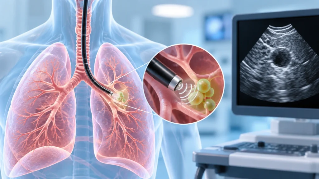
In traditional bronchoscopy, a thin flexible tube is passed through the nose or mouth into the airways to look inside the lungs. However, the bronchoscope can only see the inner surface of the airways. Many important structures, such as lymph nodes and blood vessels, lie outside the airway walls and cannot be seen directly. EBUS solves this problem by adding a small ultrasound probe at the tip of the bronchoscope. This allows the doctor to visualize these structures in real time and guide a needle to collect tissue samples safely and accurately.

What is Endobronchial Ultrasound (EBUS)?

Endobronchial Ultrasound is a specialized form of bronchoscopy that uses ultrasound imaging to examine structures around the airways. The procedure is usually called EBUS-TBNA, which stands for Endobronchial Ultrasound-guided Transbronchial Needle Aspiration.

During EBUS, a pulmonologist inserts a bronchoscope with an ultrasound probe through the nose or mouth into the windpipe and major airways. The ultrasound creates images of the airway walls, nearby lymph nodes, blood vessels, and other structures. Using these images, the doctor can guide a fine needle through the bronchoscope wall into the target area and collect cells or tissue for testing.

EBUS is different from standard bronchoscopy because it allows sampling of structures that are outside the airway. It is also different from surgical biopsy because it does not require large incisions, general anesthesia in all cases, or prolonged hospital stay. Most patients go home the same day and resume normal activities within a short time.

Why is EBUS Done?

EBUS is primarily a diagnostic and staging procedure. It helps doctors answer important questions about lung and chest conditions.

Common reasons for EBUS include:

  • Evaluating enlarged lymph nodes in the chest seen on CT scan or X-ray.
  • Diagnosing lung cancer and determining its stage.
  • Checking whether cancer has spread to lymph nodes.
  • Investigating suspicious masses near the airways.
  • Diagnosing infections such as tuberculosis involving chest lymph nodes.
  • Evaluating unexplained findings in the mediastinum, which is the central part of the chest.
  • Assessing lymph nodes in patients with lymphoma or other cancers.
  • Guiding treatment decisions such as surgery, chemotherapy, or radiation.

Why EBUS is important in lung cancer: Accurate staging is critical in lung cancer. Treatment options depend on whether the cancer is confined to the lung or has spread to lymph nodes or other areas. EBUS allows direct sampling of lymph nodes to confirm or rule out spread. This helps avoid unnecessary surgery in some patients and ensures the right treatment plan for others.

Types of EBUS Procedures

There are two main types of endobronchial ultrasound procedures, each suited for different areas of the chest.

Convex probe EBUS

  • Uses a bronchoscope with an ultrasound probe that curves outward.
  • Ideal for sampling lymph nodes along the windpipe and major airways.
  • Commonly used for lung cancer staging and mediastinal lymph node evaluation.
  • Performed through the mouth or nose into the trachea and bronchi.

Radial probe EBUS

  • Uses a small rotating ultrasound probe that can be passed through a standard bronchoscope.
  • Ideal for evaluating small nodules or lesions in the outer parts of the lung.
  • Often used to guide biopsy of peripheral lung nodules seen on CT scan.
  • Helps increase the accuracy of sampling small or hard-to-reach lesions.

In many centers, both types may be used depending on the clinical question and the location of the abnormality.

Benefits of EBUS

EBUS offers several advantages over traditional surgical methods for chest evaluation.

Key benefits include:

  • Minimally invasive with no external incisions.
  • Performed through natural airways, reducing trauma.
  • Can be done under sedation rather than general anesthesia in many cases.
  • Shorter procedure time compared to surgical biopsy.
  • Same-day discharge for most patients.
  • Lower risk of complications compared to open surgery.
  • High accuracy for sampling mediastinal lymph nodes.
  • Real-time ultrasound guidance for precise needle placement.
  • Ability to sample multiple lymph node stations in one session.
  • Reduces the need for more invasive procedures such as mediastinoscopy.

Why patients prefer EBUS: Patients often prefer EBUS because it is less painful, requires less recovery time, and avoids large scars. It also provides quick results, which helps in faster treatment planning. For elderly patients or those with other health problems, EBUS is often a safer option than surgery.

How is EBUS Performed?

The EBUS procedure is performed by a trained pulmonologist or interventional bronchoscopist in a procedure room or endoscopy suite.

Before the procedure

  • The patient is assessed for fitness and any bleeding risk.
  • Fasting is required for several hours before the procedure.
  • Medicines may be adjusted, especially blood thinners.
  • Consent is taken after explaining the procedure, benefits, and risks.
  • An intravenous line is placed for sedation and medications.

During the procedure

  • The patient is given sedation to relax and reduce discomfort.
  • Oxygen is provided through a nasal cannula or mask.
  • The bronchoscope is gently passed through the nose or mouth into the windpipe.
  • Ultrasound imaging is used to locate the target lymph nodes or masses.
  • A fine needle is passed through the bronchoscope into the target under ultrasound guidance.
  • Multiple samples may be taken from different areas if needed.
  • The samples are sent to the laboratory for analysis.

After the procedure

  • The patient is monitored in a recovery area until sedation wears off.
  • Vital signs, oxygen levels, and breathing are checked.
  • Most patients can go home the same day once stable.
  • Results are discussed once the pathology reports are available.

Preparation for EBUS

Proper preparation helps ensure the procedure is safe and successful.

Pre-procedure instructions

  • Fast for at least six to eight hours before the procedure.
  • Inform the doctor about all medicines, especially blood thinners.
  • Mention any history of bleeding problems or allergies.
  • Share details of previous lung procedures or surgeries.
  • Arrange for someone to accompany you home after the procedure.
  • Avoid driving or operating machinery on the day of the procedure.

Tests that may be done before EBUS

  • Blood tests to check hemoglobin, platelets, and clotting.
  • Chest imaging such as CT scan to plan the procedure.
  • Lung function tests if needed.
  • Heart evaluation in patients with significant heart disease.

Medication adjustments

  • Blood thinners may need to be paused or adjusted before the procedure.
  • Diabetes medicines may need dose changes due to fasting.
  • Regular medicines for blood pressure or heart conditions are usually continued.
  • Always follow the specific instructions given by the treating team.

Recovery After EBUS

Recovery from EBUS is usually quick because the procedure is minimally invasive.

Immediate recovery

  • Patients are observed for one to two hours after the procedure.
  • Throat numbness from local anesthesia may last for a short time.
  • Mild sore throat, cough, or hoarseness is common.
  • Small amount of blood-tinged sputum may occur if biopsy was done.

Home care instructions

  • Rest for the remainder of the day.
  • Avoid hot or spicy foods if the throat is sore.
  • Do not drive or make important decisions until the next day.
  • Resume normal diet as tolerated.
  • Take prescribed medicines as directed.
  • Report any severe symptoms immediately.

Risks and Complications

EBUS is generally safe, but like any medical procedure, it carries some risks.

Common minor issues

  • Sore throat.
  • Mild cough.
  • Hoarseness.
  • Small amount of blood in sputum.
  • Temporary discomfort in the chest or throat.

Less common complications

  • Bleeding from the biopsy site.
  • Infection at the sampling site.
  • Low oxygen levels during or after the procedure.
  • Reaction to sedation medicines.
  • Injury to the airway wall.

Rare but serious complications

  • Significant bleeding requiring intervention.
  • Pneumothorax, which is air leak around the lung.
  • Severe infection or abscess formation.
  • Heart rhythm problems in susceptible patients.
  • Need for emergency surgery in very rare cases.

Why complications are uncommon: EBUS is performed under direct ultrasound visualization, which reduces the risk of hitting blood vessels or vital structures. The needle used is very fine, and the procedure is done by trained specialists. Careful patient selection and monitoring further reduce risks.

Who is a Candidate for EBUS?

Not every patient with a chest abnormality needs EBUS. The decision depends on the clinical situation and imaging findings.

Good candidates for EBUS include:

  • Patients with enlarged mediastinal or hilar lymph nodes on CT scan.
  • Individuals with suspected lung cancer needing staging.
  • Patients with unexplained chest masses near the airways.
  • Those with suspected tuberculosis involving chest lymph nodes.
  • People with lymphoma or other cancers needing lymph node evaluation.
  • Patients who are not fit for surgical biopsy due to other health issues.

When EBUS may not be needed

  • Small peripheral nodules that can be reached by other biopsy methods.
  • Clearly benign findings that do not require tissue diagnosis.
  • Patients with severe bleeding risk that cannot be managed safely.
  • Situations where imaging and clinical picture are sufficient for treatment decisions.

EBUS in Lung Cancer Staging

One of the most important uses of EBUS is in lung cancer staging. Accurate staging determines whether surgery is appropriate or whether other treatments are needed.

How EBUS helps in staging

  • Samples lymph nodes in the mediastinum and around the lungs.
  • Confirms whether cancer has spread beyond the lung.
  • Helps assign the correct stage to the cancer.
  • Guides decisions about surgery, chemotherapy, or radiation.
  • Avoids unnecessary surgery in patients with advanced disease.

Why staging matters: Treatment for early-stage lung cancer may involve surgery with curative intent. In contrast, advanced-stage disease may require chemotherapy, radiation, or targeted therapy. EBUS provides the tissue confirmation needed to make these decisions accurately.

EBUS for Infections and Other Conditions

EBUS is not only used for cancer. It is also valuable in diagnosing infections and other chest conditions.

Uses in infections

  • Diagnosing tuberculosis involving chest lymph nodes.
  • Sampling lymph nodes in fungal infections.
  • Evaluating unexplained fever with enlarged lymph nodes.
  • Guiding antibiotic or anti-tuberculosis treatment based on tissue results.

Uses in other conditions

  • Evaluating lymph nodes in sarcoidosis.
  • Diagnosing lymphoma involving the chest.
  • Investigating unexplained mediastinal masses.
  • Assessing response to treatment in some cases.

Why Choose Rama Hospital for EBUS?

EBUS requires specialized equipment, advanced training, and a multidisciplinary approach. A hospital with experienced pulmonologists, modern bronchoscopy suites, and strong pathology support can ensure accurate diagnosis and safe procedures.

  • Experienced interventional pulmonologists trained in advanced bronchoscopy.
  • High-quality ultrasound bronchoscopes and imaging systems.
  • On-site pathology support for rapid and accurate analysis.
  • Comprehensive pre-procedure evaluation and post-procedure care.
  • Coordination with oncology, thoracic surgery, and infectious disease teams.
  • Emergency support in case of rare complications.

Frequently Asked Questions (FAQs)

How long does the EBUS procedure take?

The procedure typically takes 30 to 60 minutes, depending on the number of samples needed. Total time at the hospital may be longer due to preparation and recovery.

Do I need to stay in the hospital overnight?

Most patients can go home the same day after a short recovery period. Overnight stay is rarely needed unless there are complications or other medical reasons.

What are the risks of EBUS?

EBUS is generally safe. Minor issues like sore throat or mild bleeding are common. Serious complications such as significant bleeding or infection are rare but possible.

When will I get the results?

Initial findings may be available within a few days. Final pathology reports, especially for cancer or infection, may take one to two weeks depending on the tests required.

Can EBUS diagnose lung cancer?

Yes, EBUS is commonly used to diagnose and stage lung cancer by sampling lymph nodes and masses in the chest. It helps determine the extent of disease and guides treatment planning.

Is EBUS better than surgical biopsy?

For many chest conditions, EBUS is less invasive, safer, and equally accurate compared to surgical biopsy. It avoids large incisions and has a shorter recovery time. However, the best approach depends on the individual case.

Conclusion

Endobronchial Ultrasound (EBUS) is a modern, minimally invasive procedure that has transformed the diagnosis and staging of lung and chest conditions. It allows doctors to see beyond the airway walls and take precise tissue samples from lymph nodes and masses without open surgery. This leads to faster diagnosis, accurate staging, and better treatment planning, especially in lung cancer and chest infections.

For patients with enlarged lymph nodes, suspicious chest findings, or suspected lung cancer, EBUS offers a safe and effective option with quick recovery. With experienced specialists, advanced technology, and comprehensive care, hospitals can provide high-quality EBUS services that improve patient outcomes.

Rama Hospital offers advanced EBUS procedures performed by trained pulmonologists, supported by modern imaging and pathology services. Patients receive personalized care, clear communication, and timely results to guide their treatment journey with confidence.
Table of Contents

Explore More

What to Know More About Endobronchial Ultrasound (EBUS)

Find Ailments & Treatments

Search Ailments

Quickly find the information you need. Search our database to explore detailed information on various diseases and conditions, including symptoms, causes, and treatment options.

Search Treatments

Quickly find the information you need. Search our database to explore detailed information on various diseases and conditions, including symptoms, causes, and treatment options.

Recommended Reading

Video Playlist

No rows found.

Experience Healthcare the Rama Way

Emergency • OPD • Surgery • ICU Care

Need Help? Talk to our experts

Get a callback from our experts

Request a callback