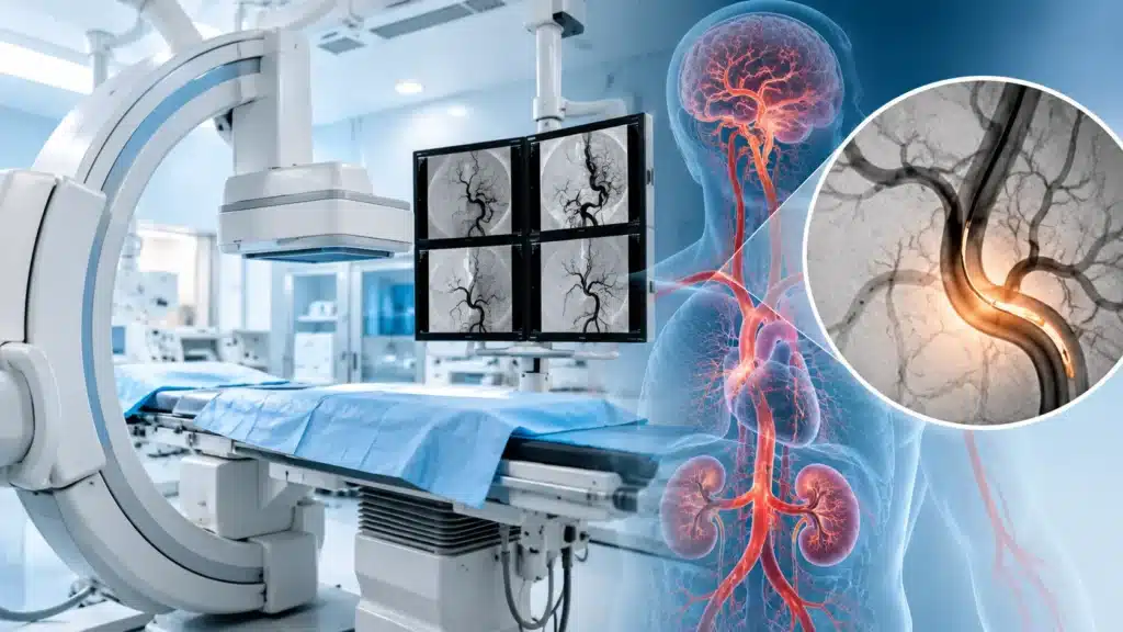
Digital Subtraction Angiography, commonly known as DSA, is an advanced imaging test that helps doctors see blood vessels in the body in great detail. It is mainly used to study arteries and veins in the brain, heart, kidneys, limbs, and other organs. Digital Subtraction Angiography is especially useful when doctors need a very clear view of blood flow, blockages, narrowing, abnormal connections, or bulges in blood vessels.
Unlike a normal X-ray, Digital Subtraction Angiography uses a special technique where images taken before and after injecting a contrast dye are compared. The computer removes or “subtracts” the background such as bones and soft tissues. This leaves a clear image of only the blood vessels. Because of this, even small changes in vessel structure or blood flow can be seen clearly.
What is Digital Subtraction Angiography (DSA)?
Digital Subtraction Angiography is a specialized X-ray imaging technique used to visualize blood vessels. A thin tube called a catheter is usually inserted into a blood vessel, often in the groin or wrist. A contrast dye is then injected through this tube. As the dye flows through the vessels, X-ray images are taken rapidly.
The computer then compares images taken before and after the dye injection. It removes the background structures such as bones, muscles, and organs. This leaves a clear, detailed image of only the blood vessels. This is why the technique is called “subtraction” angiography.
Digital Subtraction Angiography is considered one of the most accurate tests for studying blood vessels. It is often used by interventional radiologists, neurosurgeons, cardiologists, and vascular specialists. In many cases, DSA is not only a diagnostic test but also the first step in treatment, such as placing a stent, coil, or other device.
Why is DSA Done?
Digital Subtraction Angiography is done when doctors need a very clear and detailed view of blood vessels. It helps in both diagnosis and treatment planning.
Common reasons for DSA
- To detect blockages or narrowing in arteries.
- To study abnormal blood vessel connections.
- To detect aneurysms or bulges in blood vessels.
- To evaluate blood flow to the brain, heart, kidneys, or limbs.
- To plan surgery or interventional procedures.
- To guide treatment such as stent placement or embolization.
- To check blood supply before or after surgery.
- To investigate unexplained bleeding.
- To evaluate vascular malformations.
Common areas studied with DSA
- Brain and neck vessels for aneurysm, stroke, or vascular malformation.
- Heart and coronary arteries in selected cases.
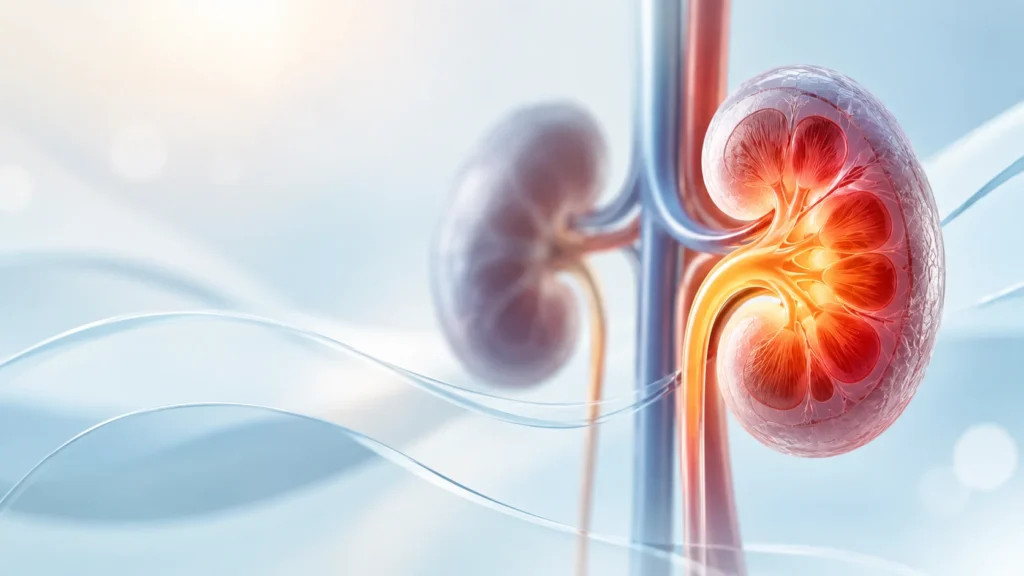
- Kidney arteries for narrowing or high blood pressure.
- Leg and arm arteries for blockage or poor circulation.
- Liver and other abdominal vessels for tumor blood supply or bleeding.
- Spinal vessels in selected neurological conditions.
Types of DSA
Digital Subtraction Angiography can be classified based on the area being studied and the purpose of the procedure.
Based on area
- Cerebral DSA, which studies blood vessels in the brain and neck.
- Coronary DSA, which studies vessels of the heart in selected cases.
- Renal DSA, which studies kidney arteries.
- Peripheral DSA, which studies vessels in the arms or legs.
- Visceral DSA, which studies vessels of the abdomen such as liver, spleen, or intestines.
- Spinal DSA, which studies vessels around the spine in selected cases.
Based on purpose
- Diagnostic DSA, which is done to identify or confirm a problem.
- Therapeutic DSA, which is done to guide treatment such as stent placement, coil placement, or embolization.
- Follow-up DSA, which is done to check results after treatment or monitor a known condition.
How Does DSA Work?
Digital Subtraction Angiography works by using contrast dye and computer image processing to create clear images of blood vessels.
Step-by-step principle
- A contrast dye is injected into the bloodstream through a catheter.
- X-ray images are taken before the dye reaches the area of interest.
- More images are taken as the dye flows through the vessels.
- The computer compares the two sets of images.
- Background structures such as bones and organs are subtracted.
- Only the blood vessels filled with dye remain visible.
- Doctors can then study the vessels in detail.
Why the subtraction technique helps
Without subtraction, bones and soft tissues can hide small vessels or subtle changes. By removing these backgrounds, DSA gives a much clearer view. This helps doctors detect small aneurysms, minor blockages, or abnormal connections that may not be visible on other tests.
Conditions Diagnosed or Treated with DSA
Digital Subtraction Angiography is used in many medical specialties because it gives detailed vascular information.
Brain and nervous system
- Brain aneurysm.
- Arteriovenous malformation or AVM.
- Stroke or blocked brain artery.
- Narrowing of neck or brain arteries.
- Vascular tumors.
- Unexplained brain hemorrhage.
Heart and chest
- Coronary artery blockage in selected cases.
- Abnormal connections between vessels.
- Evaluation before or after heart procedures.
Kidneys and abdomen
- Renal artery narrowing causing high blood pressure.
- Kidney tumor blood supply.
- Gastrointestinal bleeding.
- Liver tumors or vascular malformations.
- Splenic artery problems.
Limbs and peripheral vessels
- Blockage in leg or arm arteries.
- Poor circulation or peripheral artery disease.
- Aneurysm in limb vessels.
- Planning for bypass or stent procedures.
Preparation Before Digital Subtraction Angiography
Proper preparation helps make the procedure safe and smooth.
Before the procedure
- Follow fasting instructions given by the hospital.
- Inform the team about allergies, especially to contrast dye or iodine.
- Share history of kidney problems or previous contrast reactions.
- Inform about all medicines, especially blood thinners.
- Mention pregnancy if applicable.
- Arrange for someone to accompany you home if sedation is used.
- Follow instructions about stopping or continuing medicines.
Tests that may be done before Digital Subtraction Angiography
- Blood tests to check kidney function and clotting.
- Blood sugar test if diabetic.
- ECG or other heart tests if needed.
- Imaging such as ultrasound, CT, or MRI may already be available.
- Review of previous reports and medicines.
What to bring
- Previous reports and imaging.
- List of current medicines.
- Identification and insurance documents if applicable.
- Comfortable clothing and personal items for hospital stay if needed.
How is DSA Performed?
Digital Subtraction Angiography is usually done in a special imaging room called a cath lab or angiography suite. It is performed by an interventional radiologist or vascular specialist with a trained team.
Step-by-step procedure
- The patient is positioned on the X-ray table.
- The area for catheter entry, usually groin or wrist, is cleaned and numbed.
- A small needle is used to access the blood vessel.
- A thin tube called a catheter is guided to the target area.
- Contrast dye is injected through the catheter.
- X-ray images are taken rapidly as dye flows through vessels.
- The computer subtracts background images to show vessels clearly.
- If needed, treatment such as stent, coil, or embolization is done.
- The catheter is removed and pressure is applied to stop bleeding.
- A small bandage or closure device is used at the entry site.
Duration of procedure
- Simple diagnostic DSA may take 30 to 60 minutes.
- Complex or therapeutic DSA may take longer, sometimes 1 to 3 hours.
- Recovery and observation time may add a few hours.
Sedation and comfort
- Local anesthesia is used at the entry site.
- Mild sedation may be given to help relaxation.
- The patient is usually awake but comfortable.
- The team monitors heart rate, oxygen, and blood pressure throughout.
Benefits of DSA
Digital Subtraction Angiography offers several advantages over other imaging tests when detailed vascular information is needed.
Key benefits
- Very clear and detailed images of blood vessels.
- Ability to detect small aneurysms or subtle blockages.
- Real-time imaging during the procedure.
- Can be used for both diagnosis and treatment.
- Helps plan surgery or interventional procedures accurately.
- Allows immediate treatment such as stent or coil placement.
- Reduces need for exploratory surgery in some cases.
- Provides information that other tests may miss.
Why DSA is considered gold standard
For many vascular conditions, Digital Subtraction Angiography is considered the most accurate test because it shows vessels in real time with high detail. It is especially valuable in brain aneurysm, AVM, complex blockages, and vascular malformations where precise mapping is needed.
Risks and Safety of DSA
Like any medical procedure, Digital Subtraction Angiography has some risks, but serious complications are uncommon when done by experienced teams.
Common minor risks
- Bruising or swelling at the catheter entry site.
- Mild pain or discomfort.
- Temporary feeling of warmth when dye is injected.
- Mild headache or nausea.
- Allergic reaction to contrast dye in some patients.
Less common but serious risks
- Bleeding or hematoma at the entry site.
- Damage to the blood vessel.
- Kidney stress due to contrast dye.
- Allergic reaction or anaphylaxis in rare cases.
- Stroke or neurological complication in brain DSA, though rare.
- Infection at the entry site.
How risks are reduced
- Careful patient selection and pre-procedure testing.
- Use of the lowest possible dye dose.
- Hydration before and after the procedure.
- Monitoring during and after the procedure.
- Experienced interventional team and sterile technique.
- Clear instructions for recovery and follow-up.
Recovery After Digital Subtraction Angiography
Recovery depends on whether the procedure was diagnostic or therapeutic, and on the entry site used.
Immediately after the procedure
- The patient is monitored for a few hours.
- Pressure or a closure device is used at the entry site.
- Bed rest may be advised for a few hours.
- Fluids may be given to help flush out the dye.
- Vital signs and entry site are checked regularly.
At home
- Keep the entry site clean and dry.
- Avoid heavy lifting or strenuous activity for a few days.
- Follow instructions about medicines and activity.
- Watch for signs of bleeding, swelling, or severe pain.
- Attend follow-up appointments as advised.
When to seek urgent help
- Heavy bleeding or swelling at entry site.
- Severe pain or numbness in the limb.
- Fever or signs of infection.
- Sudden weakness, speech difficulty, or vision change.
- Severe headache or confusion.
DSA vs Other Imaging Tests
Digital Subtraction Angiography is one of several tests used to study blood vessels. Each has its own role.
Comparison pointers
- Ultrasound is non-invasive and good for basic flow assessment but less detailed.
- CT angiography is fast and good for many vessels but uses radiation and dye.
- MR angiography avoids radiation in many cases but may be less detailed for small vessels.
- Digital Subtraction Angiography gives the most detailed real-time view and allows treatment in the same sitting.
- The choice depends on the condition, urgency, and patient factors.
When is DSA Recommended?
Digital Subtraction Angiography is recommended when detailed vascular information is needed for diagnosis or treatment.
Common situations
- Suspicion of brain aneurysm or AVM.
- Stroke or blocked brain artery needing intervention.
- Unexplained bleeding in the brain or abdomen.
- Severe narrowing of kidney or limb arteries.
- Planning for stent, coil, or embolization.
- Follow-up after vascular treatment.
- Complex vascular malformations.
Why Choose Rama Hospital for DSA?
Digital Subtraction Angiography is a specialized procedure that requires expert interventional radiologists, advanced imaging equipment, and a trained support team. A hospital with experience in vascular and neuro-interventional care can make a major difference in safety and outcomes
- Experienced interventional radiology and neurosurgery teams.
- Modern cath lab and imaging technology.
- Safe use of contrast dye with kidney protection protocols.
- Ability to perform both diagnosis and treatment in one procedure.
- Intensive care support if needed for complex cases.
- Clear pre-procedure counseling and post-procedure follow-up.
Frequently Asked Questions (FAQs)
Is DSA painful?
Most patients feel only mild discomfort at the catheter entry site. Local anesthesia is used to numb the area, and mild sedation may be given. The injection of dye may cause a brief warm feeling but is usually not painful.
How long does a DSA procedure take?
A simple diagnostic DSA may take 30 to 60 minutes. If treatment such as stent or coil placement is done at the same time, it may take 1 to 3 hours. Recovery and observation add a few more hours.
Is DSA safe?
Digital Subtraction Angiography is generally safe when performed by experienced teams. Like any procedure, it has some risks such as bleeding, allergic reaction, or kidney stress from dye. These risks are minimized with proper preparation and monitoring.
Do I need to stay in hospital after DSA?
Many patients go home the same day after a simple diagnostic DSA. If treatment was done or if the patient has other health issues, an overnight stay may be advised. Your doctor will give clear instructions based on your case.
Can DSA treat problems, not just diagnose?
Yes, DSA can be used for both diagnosis and treatment. During the same procedure, doctors may place a stent, coil, or perform embolization to treat blockages, aneurysms, or abnormal vessels.
What should I do before DSA?
Follow fasting instructions, inform the team about allergies and medicines, and arrange for someone to accompany you if needed. Blood tests and review of kidney function may be done before the procedure.
When will I get the results?
In many cases, the doctor can explain the findings soon after the procedure. If treatment was done, a detailed report and follow-up plan will be shared. Complex cases may need more time for full analysis.
Conclusion
Digital Subtraction Angiography (DSA) is a highly accurate imaging test that gives detailed views of blood vessels in the body. It is used to diagnose and sometimes treat conditions such as aneurysms, blockages, vascular malformations, and abnormal bleeding. Because it provides real-time, high-detail images, DSA is often considered the gold standard for many vascular problems.
The procedure is generally safe when done by experienced teams with proper preparation and monitoring. Recovery is usually quick for diagnostic DSA, while therapeutic DSA may need more observation. With the right care, DSA can help doctors make accurate diagnoses and deliver targeted treatments that improve outcomes.
Rama Hospital can provide expert evaluation, advanced DSA imaging, and coordinated care for vascular and neurological conditions. From preparation to recovery, the focus is on patient safety, accurate diagnosis, and effective treatment.