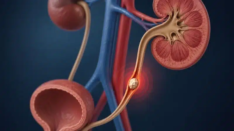
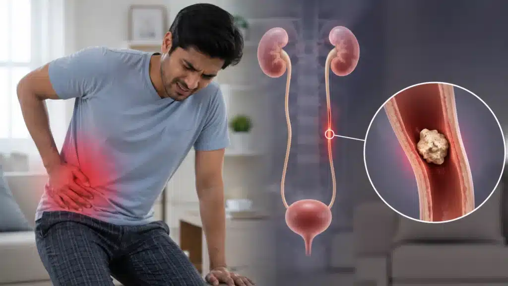
A stone stuck in the ureter, the narrow tube connecting your kidney to your bladder, is one of the most painful experiences a person can go through. The pain is sudden, intense, and completely disruptive. And for many people, the natural question that follows the diagnosis is, “How do we get it out?”
Ureteroscopy (URS) is one of the most effective and widely used answers to that question.
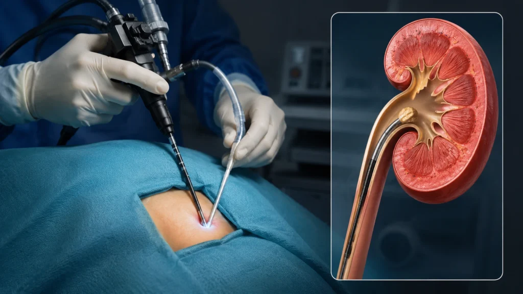
It is a minimally invasive procedure where a thin, lighted instrument called a ureteroscope is passed through the body’s natural urinary passage, through the urethra, into the bladder, and up into the ureter or kidney to directly visualize and treat the stone. No cuts. No external incisions. No open surgery.
Combined with holmium laser technology, ureteroscopy can precisely fragment even stubborn stones and remove them completely, often in a single session.
What is ureteroscopy (URS)?
Ureteroscopy is a minimally invasive urological procedure that uses a thin, telescope-like instrument, called a ureteroscope, to directly visualize the inside of the ureter and kidney collecting system.
Here’s how the procedure works:
- A thin telescope called a ureteroscope is passed through the urethra and bladder.
- The scope is carefully guided into the ureter, the tube that carries urine from the kidney to the bladder.
- For kidney stones, a flexible scope can be advanced directly into the kidney.
- The stone is located using a tiny camera attached to the scope.
- A Holmium laser is used to break the stone into small fragments or fine dust.
- Stone fragments are either removed with a small basket or passed naturally in the urine.
- The entire procedure is performed without any external cuts or stitches.
What are the types of ureteroscopy?
The type of ureteroscopy used depends on the size and location of the stone, helping ensure the safest and most effective treatment for each patient.
Rigid Ureteroscopy
- Uses a straight, rigid scope.
- Best for stones in the lower and middle ureter.
- Commonly used for ureteric stones.
Semi-Rigid Ureteroscopy
- Slightly more flexible than a rigid scope.
- Can reach the upper ureter.
- Useful for stones located higher in the urinary tract.
Flexible Ureteroscopy (RIRS)
- Uses a highly flexible scope.
- Can reach all areas of the kidney and ureter.
- Ideal for kidney stones and difficult-to-reach stones.
- Forms the basis of RIRS (Retrograde Intrarenal Surgery).
What conditions can be treated with ureteroscopy?
Ureteroscopy is primarily known for kidney and ureter stone treatment, but its applications extend to a range of other urological conditions where direct visualization and treatment of the urinary tract is needed.
Ureteral Stones (Ureteral Calculi)
- The most common indication for ureteroscopy.
- Treats stones at any level of the ureter: lower, mid, and upper.
- Particularly the preferred treatment for lower and mid-ureteral stones, outperforming ESWL in stone-free rates at these locations.
- Single-session stone-free rates are excellent, one of URS’s greatest strengths.
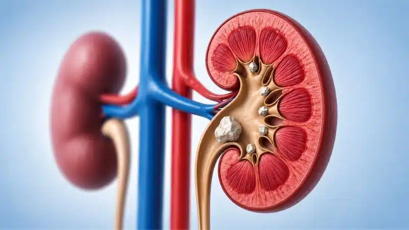
Kidney Stones (Renal Calculi)
- Using flexible ureteroscopy, stones within the kidney can be treated without any puncture.
- Ideal for kidney stones up to 2–2.5 cm.
- Particularly effective for lower pole stones, hard stones resistant to ESWL, and stones in calyceal diverticula.
- When performed for intrarenal stones, this is specifically called RIRS (Retrograde Intrarenal Surgery).
Ureteral Strictures
- Narrowing of the ureter, from scarring, previous surgery, radiation, or recurrent stone disease, causes urinary obstruction and can damage kidney function.
- Ureteroscopy allows direct visualization of the stricture.
- Endoureterotomy, cutting the stricture with a laser through the ureteroscope, can open up the narrowing and restore normal urine flow.
Transitional Cell Carcinoma (TCC) of the Ureter and Renal Pelvis
- Small, low-grade urothelial tumors in the ureter or kidney collecting system can be biopsied and, in selected cases, treated with laser ablation through the ureteroscope.
- Ureteroscopy is also the primary tool for surveillance biopsy of the upper urinary tract in patients with known or suspected urothelial cancer.
- Performed under careful oncological guidance at Rama Hospital.
Haematuria Evaluation (Blood in Urine)
- When blood in the urine has no clear cause on CT or ultrasound, diagnostic ureteroscopy allows direct inspection of the entire ureter and kidney collecting system.
- Identifies the source of bleeding, whether from a lesion, vessel, or subtle tumor, that imaging alone may miss.
Ureteral Polyps and Benign Lesions
- Benign fibroepithelial polyps of the ureter can cause obstruction and hematuria.
- Ureteroscopy allows direct visualization and laser resection of these lesions.
Foreign Bodies in the Ureter
- Occasionally, ureteral stents fragment or become encrusted and cannot be removed with simple cystoscopy.
- Ureteroscopy allows direct access to retrieve broken stent pieces or encrusted foreign bodies anywhere in the ureter or kidney.
Fungal Balls (Bezoars) in the Ureter
- In immunocompromised patients, fungal infections can form obstructing masses within the ureter.
- Ureteroscopy allows direct access and removal of these collections.
Ureteropelvic Junction (UPJ) Obstruction (Endopyelotomy)
- In selected patients with UPJ obstruction, narrowing at the junction between the kidney and ureter, a laser endopyelotomy can be performed through the flexible ureteroscope.
- Cuts the obstructing segment from inside, relieving obstruction without open surgery.
Who needs ureteroscopy treatment?
Ureteroscopy is recommended when a kidney stone, ureteral stone, or urinary tract condition requires direct treatment and is unlikely to improve on its own. Ureteroscopy is a safe, minimally invasive procedure that allows doctors to diagnose and treat many urinary tract conditions without any external cuts or incisions.
You may be a good candidate for ureteroscopy if:
- You have a ureteral stone causing pain, blockage, or urinary symptoms.
- Your stone is larger than 6–7 mm and is unlikely to pass naturally.
- You have a kidney stone up to 2–2.5 cm that needs treatment.
- Your stone is hard and resistant to shock wave therapy (ESWL).
- Previous ESWL treatment has not been successful.
- You have a narrowing (stricture) in the ureter causing obstruction.
- You are pregnant and have a symptomatic stone requiring treatment.
- You are taking blood-thinning medications and need a low-bleeding-risk procedure.
- You have a single functioning kidney, so preserving kidney function is especially important.
- You have blood in the urine (hematuria) that needs further investigation.
- Your doctor suspects a tumor in the ureter or kidney collecting system, and a biopsy is required.
What are the causes and risk factors?
Most conditions treated with ureteroscopy, especially kidney stones and ureter stones, develop gradually over time. Understanding what causes them can help reduce the risk of future problems.
Common causes of kidney and ureter stones
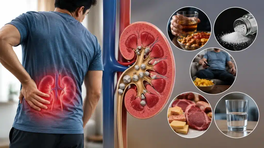
- Not drinking enough water – The most common cause. Concentrated urine allows minerals to stick together and form stones.
- Unhealthy dietary habits – Excess salt, red meat, sugary foods, and oxalate-rich foods such as spinach, nuts, tea, and chocolate can increase stone formation.
- Metabolic disorders – Certain conditions increase calcium, uric acid, or oxalate levels in the urine.
- Urinary tract infections (UTIs) – Some infections can change urine chemistry and lead to stone formation.
- Structural urinary tract problems – Conditions that slow or block urine flow can increase the risk of stones.
- Low citrate levels in urine – Citrate naturally helps prevent stones from forming. Low levels make stone formation more likely.
Common causes of ureteral strictures
A ureteral stricture is a narrowing of the ureter that can block urine flow. Common causes include:
- Previous kidney or ureter stones causing scarring.
- Past urological procedures or surgeries.
- Radiation treatment to the pelvic area.
- Chronic inflammation around the ureter.
- Congenital narrowing present from birth.
- Pressure from nearby enlarged lymph nodes or tumors.
Risk factors for kidney and ureter stones
Your risk may be higher if you:
- Do not drink enough water regularly.
- Have a family history of kidney stones.
- Have had kidney stones before.
- Are overweight or obese.
- Live in a hot climate or sweat heavily.
- Follow a high-salt or high-protein diet.
- Lead a sedentary lifestyle.
- Have conditions such as gout, hyperparathyroidism, Crohn’s disease, or renal tubular acidosis.
- Are between 30 and 60 years of age.
- Are male, although kidney stone rates are increasing among women as well.
Diagnosis before ureteroscopy
Before recommending ureteroscopy, your doctor will perform a detailed evaluation to understand the exact cause of your symptoms and plan the safest and most effective treatment.
These tests help your doctor understand your condition clearly, choose the right treatment approach, and ensure ureteroscopy is performed as safely and effectively as possible.
Imaging Tests
Non-Contrast CT KUB (Most Accurate Test for Stones)
- The gold standard for diagnosing kidney and ureter stones.
- Shows the exact size, number, and location of stones.
- Helps determine stone hardness, which guides laser treatment planning.
- Detects urinary blockage and any abnormalities in the urinary tract.
Ultrasound KUB
- A safe, radiation-free scan.
- Useful for detecting kidney stones and kidney swelling (hydronephrosis).
- Often used as an initial screening test.
X-Ray KUB
- Helps identify certain types of kidney and ureter stones.
- Commonly used for treatment planning and follow-up monitoring.
CT Urogram
- Provides detailed images of the kidneys, ureters, and bladder.
- Helpful when a ureteral blockage, narrowing, or tumor is suspected.
MRI Urogram
- Used when a CT scan is not suitable, such as during pregnancy.
- Offers detailed images of the urinary tract and surrounding tissues.
Laboratory Tests
Urine Analysis and Urine Culture
- Checks for infection, blood, or other abnormalities in the urine.
- Any infection is treated before the procedure.
Blood Tests
- Assess kidney function and overall health.
- Check blood counts and clotting ability before surgery.
Urine Cytology
- May be recommended if a urinary tract tumor is suspected.
- Looks for abnormal cells in the urine.
24-Hour Urine Test
- Often advised for patients with recurrent kidney stones.
- Helps identify factors contributing to stone formation.
Stone Analysis
- If you have passed a stone before, it may be analyzed to determine its composition.
- Helps create a personalized plan to prevent future stones.
Treatment procedure: how is ureteroscopy done?
One of the biggest advantages of ureteroscopy is that it treats kidney and ureter stones without any cuts or incisions. Using a thin camera and advanced laser technology, the stone can be located, broken, and removed through the natural urinary passage.
Before the Procedure
Before treatment, your doctor will perform a detailed evaluation to ensure the procedure is safe and effective.
- Blood tests, urine tests, and imaging scans.
- Treatment of any existing urinary tract infection.
- Fasting for a few hours before the procedure.
- Anesthesia assessment.
- In some cases, a temporary stent may be placed before surgery to make the procedure easier.
- Preventive antibiotics are given before treatment.
During the Procedure
Step 1: Reaching the Stone
- A thin ureteroscope is passed through the urethra and bladder into the ureter.
- No cuts or external incisions are required.
- The urinary tract is carefully examined as the scope advances.
Step 2: Locating the Stone
- The stone is identified using a high-definition camera.
- Its size, location, and position are assessed.
- For kidney stones, a flexible scope can reach different parts of the kidney.
Step 3: Breaking the Stone
- A Holmium laser is used to break the stone into smaller pieces.
- Tiny fragments may pass naturally through urine.
- Larger fragments can be removed using a special basket device.
Step 4: Confirming Stone Clearance
- The treated area is rechecked to ensure the stone has been completely cleared.
- Imaging guidance may be used to confirm successful treatment.
Step 5: Stent Placement
- A temporary Double J (DJ) stent is often placed.
- This helps urine flow freely and reduces swelling during healing.
- The stent is usually removed within 1–2 weeks.
After the Procedure
Recovery is usually quick, and most patients return home the same day or the next morning.
- Recovery monitoring after anesthesia.
- Mild discomfort, burning during urination, or blood-tinged urine may occur for a few days.
- Walking and light activities can usually resume within 24 hours.
- Medications and aftercare instructions are provided before discharge.
- Follow-up visits ensure proper healing and complete stone clearance.
What are the benefits of ureteroscopy?
Ureteroscopy (URS) is a minimally invasive procedure that offers an effective and reliable solution for many kidney and ureteral stones. Because it uses the body’s natural urinary pathway, recovery is usually quick and comfortable.
With its high success rates, quick recovery, and minimally invasive approach, ureteroscopy (URS) has become one of the most commonly used treatments for kidney and ureteral stones.
Some key benefits include:
- No cuts or incisions: the procedure is performed through the natural urinary tract.
- High stone clearance rates, often in a single treatment session.
- Effective for stones located in the upper, middle, or lower ureter.
- Works well on all types of stones, including hard stones that may not respond to shock wave treatment.
- Allows direct removal of stone fragments, reducing the chance of leftover pieces.
- Can treat stones, ureteral narrowing (strictures), and other urinary tract problems during the same procedure.
- Short hospital stay, with many patients going home within 24 hours.
- Faster recovery, with most people returning to normal activities within a few days.
- Considered a safe option during pregnancy when stone treatment is necessary.
- Suitable for many patients who take blood-thinning medications.
- Helps protect kidney function, especially in people with a single functioning kidney.
- Can be used to diagnose and treat certain urinary tract conditions during the same procedure.
- In selected cases, the procedure can be performed with minimal or no radiation exposure.
What are the risks and complications of ureteroscopy?
Millions of patients undergo ureteroscopy successfully every year. Although complications are uncommon, knowing what to expect after the procedure is an important part of your treatment journey.
Common and Temporary Side Effects
These are usually mild and improve within a few days:
- Blood in the urine for a short period after the procedure.
- Burning or discomfort while urinating.
- Frequent or urgent urination.
- Mild pain or aching in the side or lower back.
- Stent-related discomfort, such as bladder irritation, urgency, or a pulling sensation if a Double J (DJ) stent has been placed.
Less Common Complications
- Urinary tract infection (UTI) that may require antibiotics.
- Residual stone fragments that may need an additional procedure.
- Minor injury to the ureter, which is uncommon and usually heals with proper treatment.
- Steinstrasse (“stone street”), where small stone fragments temporarily collect in the ureter and may need monitoring or treatment.
Rare but Serious Complications
- Ureteral stricture, where scar tissue narrows the ureter over time.
- Severe infection (sepsis), which requires urgent medical treatment.
- Significant ureteral injury, a very rare complication with modern techniques and experienced surgeons.
Tips for better recovery after ureteroscopy
Good hydration, timely follow-ups, and healthy habits are the best ways to recover well and reduce the risk of future kidney stones.
Most people recover quickly after ureteroscopy and can return to their normal routine within a few days. Following these simple tips can help ensure a smooth recovery:
- Drink plenty of water (2.5–3 liters daily) to flush out stone fragments and support healing.
- Take all prescribed medicines as directed by your doctor.
- Rest for the first 1–2 days and avoid heavy lifting or strenuous exercise.
- Mild blood in the urine is normal for a few days after the procedure.
- A ureteral stent may cause temporary discomfort, frequent urination, or urgency until it is removed.
- Do not miss your stent removal appointment.
- Follow your kidney stone prevention plan, including dietary and lifestyle changes.
- Attend all follow-up appointments and scans to confirm complete stone clearance.
- Contact your doctor immediately if you develop fever, severe pain, difficulty urinating, or heavy bleeding.
- Report any persistent symptoms that continue after stent removal.
Why choose Rama Hospital for ureteroscopy?
Choosing the right hospital can make a big difference in your treatment experience and recovery. Here’s why patients trust Rama Hospital for ureteroscopy:
- Experienced urologists with expertise in simple and complex stone treatments.
- Advanced rigid and flexible ureteroscopy technology.
- Modern Holmium laser for precise stone fragmentation.
- High stone clearance rates in a single procedure.
- Personalized kidney stone prevention and follow-up care.
- Comprehensive diagnosis, treatment, and recovery support under one roof.
- Short hospital stay with faster return to daily activities.
- Multidisciplinary team for complex urological conditions.
- Patient-focused care with clear guidance at every step.
- Dedicated support for a smooth and comfortable recovery.
When should you see a doctor for ureteroscopy treatment?
Early medical attention can help prevent complications, protect kidney function, and ensure you receive the right treatment at the right time.
- You have severe pain in your side, back, or lower abdomen that comes and goes in waves.
- You notice blood in your urine.
- You have nausea or vomiting along with kidney stone pain.
- Urination becomes painful, difficult, or causes burning.
- Your previous kidney stone symptoms have returned.
- A scan has detected a kidney stone or ureter stone, even if you have no symptoms yet.
- Your stone has not passed despite medication and hydration.
- You have been told your stone is large, hard, or difficult to treat.
- You experience unexplained blood in the urine.
- You have a ureter blockage or recurrent urinary infections.
Frequently Asked Questions (FAQs)
What is the difference between ureteroscopy and ESWL?
ESWL is completely non-invasive; shock waves are delivered from outside the body with no instruments inserted. It works best for smaller, softer kidney stones in favorable locations. Ureteroscopy involves passing a thin scope through the natural urinary passage and uses a Holmium laser to directly fragment and remove stones, achieving higher stone-free rates for virtually all ureteral stones and many kidney stones, working on all stone types regardless of hardness. For most ureteral stones, ureteroscopy is the preferred treatment.
Is ureteroscopy painful?
The procedure is performed under spinal or general anesthesia; you feel nothing during it. Afterwards, mild burning during urination, some blood in the urine, and a dull flank ache for a few days are normal, managed with prescribed medications. Most patients find these symptoms very manageable and are comfortable within 2 to 3 days.
How long does a ureteroscopy procedure take?
Most ureteroscopy procedures take between 30 and 90 minutes depending on the stone’s size, location, and complexity. Your surgeon at Rama Hospital will give a more specific estimate based on your imaging.
Will I need a stent after ureteroscopy?
In most cases, yes, a Double J ureteral stent is placed at the end of the procedure. It keeps the ureter open while swelling settles and ensures safe kidney drainage during recovery. Some urinary frequency, urgency, and mild discomfort are expected while the stent is in. It is removed in a quick, simple outpatient procedure, usually 1 to 2 weeks after ureteroscopy, after which these symptoms resolve.
What are the stone-free rates with ureteroscopy?
For ureteral stones, single-session stone-free rates with ureteroscopy are very high, typically 90 to 95%, among the best of any stone treatment available. For intrarenal stones up to 2–2.5 cm, stone-free rates are in the 85 to 95% range depending on stone size and location. Your surgeon will give you a realistic expectation based on your specific stone characteristics.
Is ureteroscopy safe during pregnancy?
Yes, ureteroscopy is considered the safest surgical option for symptomatic ureteral stones during pregnancy. It avoids radiation exposure (unlike CT scanning), and the Holmium laser is safe for the urinary tract. Our urology team works closely with our obstetrics team at Rama Hospital to manage pregnant patients with stone disease safely and effectively.
Can ureteroscopy treat a ureteral stricture at the same time as a stone?
Yes, this is one of the significant advantages of ureteroscopy. If a ureteral stricture is identified during the procedure, it can be treated simultaneously using laser endoureterotomy, cutting the stricture from inside, without needing a separate procedure. Your surgeon will plan for this if preoperative imaging suggests a stricture.
How soon can I return to work after ureteroscopy?
Most patients return to desk-based work within 3 to 5 days. Physically demanding work and vigorous exercise are typically resumed after 1 to 2 weeks, subject to your surgeon’s clearance. Recovery after ureteroscopy is significantly faster than after more invasive procedures like PCNL.
What happens if my stone doesn’t clear completely with ureteroscopy?
While single-session stone-free rates for ureteroscopy are high, very large or complex stones may occasionally require a second session. Your surgeon will assess stone clearance with follow-up imaging at 4 to 6 weeks. If fragments remain, options include a repeat ureteroscopy, ESWL for residual fragments, or conservative management if the fragments are small enough to pass naturally.
Conclusion
Ureteroscopy has transformed the treatment of ureteral and kidney stones, turning what once required open surgery or prolonged hospital stays into a precise, minimally invasive procedure with same-day or next-day discharge and a rapid return to normal life. Beyond stones, ureteroscopy plays a vital diagnostic and therapeutic role in managing ureteral strictures, urothelial lesions, and unexplained hematuria, making it one of the most versatile tools in modern urology.
At Rama Hospital, our experienced urology team combines deep procedural expertise with the latest generation rigid and flexible ureteroscopes and Holmium laser systems, delivering consistently high stone-free rates, comprehensive stone management, and genuine patient-centered care at every step.
Whether you have a painful ureteral stone, a kidney stone that has failed other treatments, or a urological condition that needs direct ureteral assessment, our team is ready to help.
Book your consultation with the urology specialists at Rama Hospital today and take the first step toward complete, lasting relief.